Pruritus is a feature of which of the following conditions?
Steroids are used in the Rx of the following diseases EXCEPT:
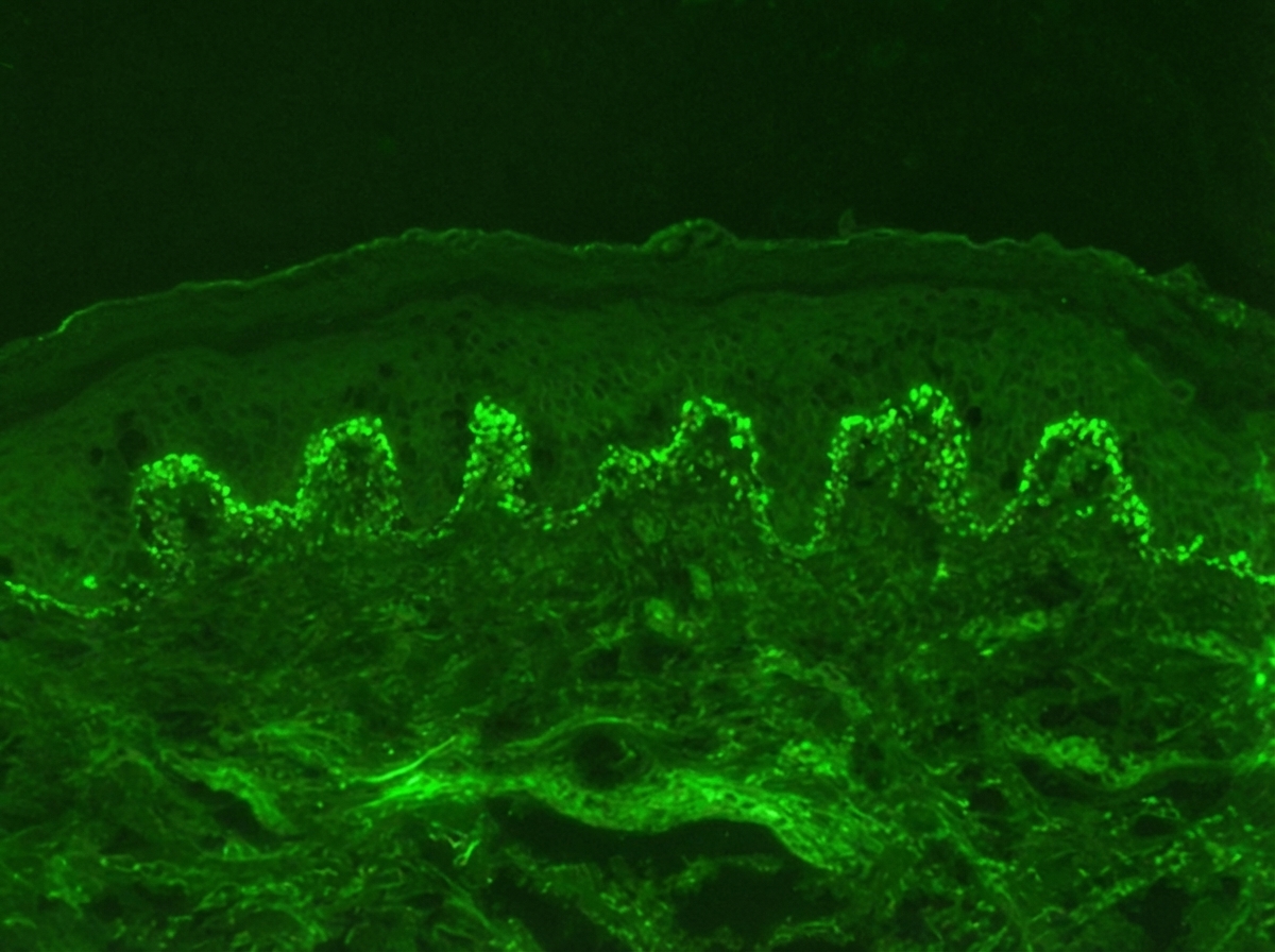
Identify the diagnosis based on the dermatology immunofluorescence (IF) image provided.

A 25-year-old patient presents with chronic itchy, erythematous skin lesions on the flexural areas that have been recurring since childhood. The patient has a family history of asthma. Which of the following is the most important diagnostic criterion for the most likely diagnosis?
"Isomorphic response" can be a feature of the following except
Which of the following is NOT a feature of atopic dermatitis?
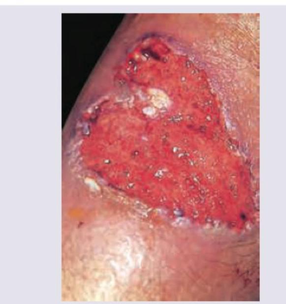
The following lesion appears on the leg of a patient of ulcerative colitis. All are useful in management except:

Patient on anti-TB drugs develops tender nodules on shins. Most likely diagnosis is:
Most common precipitant of contact dermatitis is?
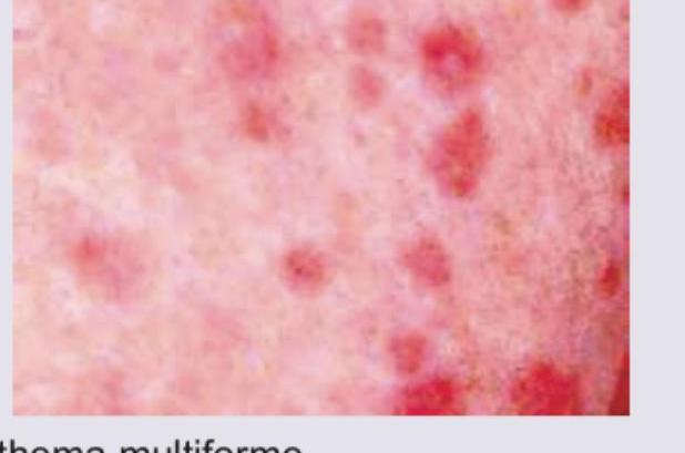
Identify the lesion: (Recent NEET Pattern 2016-17)

Explanation: ***Bullous pemphigoid*** - **Pruritus**, often severe, is a common and early symptom of bullous pemphigoid, often preceding the appearance of skin lesions. - The disease involves autoantibodies against **hemidesmosomal proteins** (BPAG1, BPAG2), leading to subepidermal blister formation. *Pemphigus foliaceous* - This condition is characterized by **superficial blistering** and erosions, but **pruritus is typically mild or absent**. - Blisters form in the **granular layer of the epidermis** due to autoantibodies against desmoglein 1. *Pemphigus vulgaris* - Patients with pemphigus vulgaris present with **flaccid blisters and erosions**, mainly affecting the skin and mucous membranes, but **pruritus is not a prominent feature**. - The disease involves intraepidermal blistering caused by autoantibodies targeting **desmoglein 3 (and sometimes desmoglein 1)**. *None of the options* - This option is incorrect, as **pruritus is a characteristic symptom of bullous pemphigoid**.
Explanation: ***Chronic fungal infection*** - **Steroids are absolutely contraindicated** in **chronic fungal infections** as they **suppress cell-mediated immunity**, leading to worsening of the infection and potential dissemination. - Corticosteroids promote fungal growth and can convert a localized infection into a systemic, life-threatening condition. - This is the **clearest contraindication** among the options. *Pemphigus vulgaris* - **Pemphigus vulgaris** is an **autoimmune blistering disease** where **high-dose systemic steroids are the first-line treatment**. - Corticosteroids (1-2 mg/kg/day of prednisolone) are essential for controlling autoantibody production and preventing life-threatening complications. - **Steroids are clearly indicated**, not contraindicated. *Erythema multiforme* - **Erythema multiforme** is typically a **self-limiting condition** managed primarily with **supportive care** (antipyretics, antihistamines, topical care). - **Systemic steroids are generally NOT recommended** as standard treatment and their use remains **controversial**. - However, in very rare severe cases with extensive mucosal involvement, some clinicians may consider a short course, making this **not an absolute contraindication** like fungal infections. *Contact dermatitis* - **Contact dermatitis** is commonly treated with **topical corticosteroids** as first-line therapy to reduce inflammation and pruritus. - In severe, widespread cases, a short course of **oral steroids** may be prescribed. - **Steroids are clearly indicated** for this condition.
Explanation: ***Dermatitis herpetiformis*** - The immunofluorescence image shows **granular IgA deposits** at the **dermal papillae region**, which is characteristic of dermatitis herpetiformis. - This condition is strongly associated with **celiac disease** and presents with intensely pruritic papules and vesicles. *Pemphigus vulgaris* - Immunofluorescence in pemphigus vulgaris typically shows a **fishnet pattern** of IgG deposits throughout the **epidermis**, reflecting antibodies against desmoglein 3 and 1. - This pattern is an intercellular deposition, not granular at the dermal papillae. *Pemphigus foliaceus* - Similar to pemphigus vulgaris, pemphigus foliaceus also exhibits **intercellular IgG deposits** in the epidermis, but it is usually more superficial, targeting desmoglein 1. - The image does not show this intercellular epidermal staining. *Bullous pemphigoid* - Bullous pemphigoid is characterized by **linear IgG and C3 deposits along the dermal-epidermal junction** (basement membrane zone). - The image distinctly shows granular IgA, not linear IgG/C3, and specifically in the dermal papillae.
Explanation: ***Chronic pruritic eczema with typical morphology and distribution*** - The patient presents with **chronic**, **itchy**, **erythematous lesions** on the **flexural areas** (e.g., antecubital and popliteal fossae), characteristic of **atopic dermatitis** (eczema). - The **recurrence since childhood** and the typical distribution represent the **major diagnostic criteria** based on clinical morphology and distribution. - **Clinical presentation with typical morphology** is the **primary diagnostic criterion** according to Hanifin and Rajka criteria. *Elevated serum IgE levels* - While **elevated serum IgE** is often associated with atopic dermatitis, it is a **minor criterion** and a **laboratory finding**, not a primary diagnostic feature. - It reflects an **atopic predisposition**, but **clinical morphology and distribution** remain the most important diagnostic factors. *Personal or family history of atopy* - A **family history of asthma** (an atopic condition) is a **minor criterion** that supports the diagnosis of atopic dermatitis. - However, this is a **predisposing/supporting factor**, not as important as the characteristic clinical morphology and distribution. *Early age of onset (before 2 years)* - While atopic dermatitis often begins in **infancy or early childhood**, this is a **minor criterion** in the diagnostic framework. - The question states symptoms **recurring since childhood** but onset timing is less diagnostically important than the characteristic **clinical presentation** with typical morphology and distribution.
Explanation: ***Tinea*** - The **isomorphic response (Koebner phenomenon)** refers to the development of new skin lesions in areas of trauma due to an immunological process. - This phenomenon is **not typically seen in tinea** (fungal infections). - While tinea can spread to new areas, this occurs through **direct fungal inoculation and contact spread**, not through the true Koebner mechanism. *Warts* - **Warts** caused by human papillomavirus (HPV) can exhibit the **isomorphic response**. - Trauma to the skin can lead to **viral inoculation** in that area, resulting in new wart formation along lines of trauma. - This is a well-recognized example of Koebner phenomenon in viral infections. *Molluscum contagiosum* - **Molluscum contagiosum** (poxvirus infection) can demonstrate the **isomorphic response**. - **Scratching or rubbing** can spread the virus to new areas through autoinoculation. - New lesions develop along the lines of trauma, consistent with Koebner phenomenon. *Psoriasis* - **Psoriasis** is the **classic and most well-known** condition exhibiting the isomorphic response or Koebner phenomenon. - New psoriatic plaques appear in areas of **skin injury** (scratches, cuts, burns, surgical incisions, friction). - Seen in approximately **25-50%** of psoriasis patients.
Explanation: ***Darier's Sign*** - **Darier's sign** is characteristic of **urticaria pigmentosa** (cutaneous mastocytosis), where rubbing a skin lesion causes the formation of an urticarial wheal due to mast cell degranulation - It is **not associated** with the pathogenesis or clinical presentation of **atopic dermatitis** *Dennie-Morgan fold* - **Dennie-Morgan folds** are extra folds or lines in the skin just below the lower eyelids - They are a common clinical sign observed in patients with **atopic dermatitis**, often linked to chronic inflammation and allergic reactions affecting the skin around the eyes *Hertoghe's sign* - **Hertoghe's sign** refers to the thinning or absence of the lateral third of the eyebrows - This sign is often seen in individuals with **atopic dermatitis**, as well as in other conditions like hypothyroidism *Hyperlinearity of palms* - **Hyperlinearity of palms** refers to the exaggerated creases and lines on the palms of the hands - This is a common **stigmata of atopy** and is frequently observed in patients with **atopic dermatitis**, reflecting the underlying predisposition to skin dryness and altered epidermal barrier function
Explanation: ***Sulfapyridine*** - The image shows **pyoderma gangrenosum**, a painful ulcerative skin condition often associated with inflammatory bowel disease like ulcerative colitis. Among the given options, **sulfapyridine** has the **least established role** in pyoderma gangrenosum management. - **Sulfapyridine** is an inactive component of **sulfasalazine** and primarily acts as an **antibacterial agent**. While sulfasalazine has been reported in some PG cases, sulfapyridine alone is not a recognized treatment for the inflammatory, non-infectious nature of pyoderma gangrenosum. - Unlike the other options which have well-established roles, sulfapyridine lacks strong evidence for efficacy in PG. *Steroids* - **Corticosteroids** (oral or topical) are the **first-line treatment** for pyoderma gangrenosum due to their potent anti-inflammatory and immunosuppressive effects. - They help to reduce the inflammation and promote healing of the painful ulcers. *Procto-colectomy* - In cases of severe, refractory pyoderma gangrenosum associated with ulcerative colitis, **colectomy** can be a **definitive treatment** as it removes the underlying inflammatory trigger. - This surgical intervention is considered when medical therapies are unsuccessful or when the colonic disease itself necessitates surgery. *Infliximab* - **Infliximab**, a **TNF-alpha inhibitor**, is a biologic agent effective in treating both ulcerative colitis and pyoderma gangrenosum. - It is used in cases that are refractory to steroids or when patients cannot tolerate steroid therapy.
Explanation: ***Erythema nodosum*** - Erythema nodosum is a common **cutaneous adverse drug reaction** to anti-TB medications, presenting with **tender, erythematous nodules** typically on the shins. - It is a form of **panniculitis** (inflammation of subcutaneous fat) specifically associated with various triggers, including infections and drugs, making it highly probable in this context. *Sweet syndrome* - Sweet syndrome (acute febrile neutrophilic dermatosis) presents with **tender, erythematous plaques and nodules** often associated with fever and leukocytosis. - While it can be drug-induced, it typically involves a more widespread skin eruption and prominent systemic symptoms like **fever**, which are not specified here. *Panniculitis* - Panniculitis is a general term for **inflammation of the subcutaneous fat**, and erythema nodosum is a type of panniculitis. - This option is too broad; while accurate, "Erythema nodosum" is the **most specific and likely diagnosis** given the patient’s presentation in the context of anti-TB drug use. *Erythema multiforme* - Erythema multiforme is characterized by **target lesions** (concentric rings of erythema and edema) and often involves mucous membranes. - The description of **tender nodules on shins** does not fit the characteristic morphology of erythema multiforme.
Explanation: ***Nickel*** - **Nickel** is the most frequent cause of **allergic contact dermatitis**, commonly found in jewelry, belt buckles, and zippers. - Exposure leads to a **Type IV hypersensitivity reaction**, characterized by erythema, itching, and vesiculation. *Gold* - While gold can cause contact dermatitis, it is **far less common** than nickel allergy. - Reactions to gold are often seen with prolonged skin contact, such as with jewelry. *Silver* - **Silver** is a **rare cause** of allergic contact dermatitis. - Allergic reactions to silver are typically observed in individuals with extensive exposure, such as jewelers. *Iron* - **Iron** is **not a common precipitant** of contact dermatitis. - Allergic reactions to iron are exceedingly rare, as iron is an essential element found naturally in the body.
Explanation: ***Erythema multiforme*** - The image displays characteristic **targetoid lesions** with multiple concentric rings of color (erythema, edema, pallor), typical of **erythema multiforme**. - These lesions often appear suddenly, symmetrically, and commonly on the extremities, often triggered by infections (e.g., **herpes simplex virus**) or medications. *Gianotti-Crosti syndrome* - Characterized by **monomorphic, flesh-colored to erythematous papules** and papulovesicles, often on the cheeks, buttocks, and extensor surfaces of the limbs. - This condition is typically observed in **children** after viral infections and does not usually present with target lesions. *Pityriasis rosea* - Starts with a single **"herald patch,"** followed by smaller, oval, pinkish-red patches with fine scales, often arranged in a **"Christmas tree pattern"** on the trunk. - The morphology of the lesions in the image, specifically the targetoid appearance, is not consistent with pityriasis rosea. *Acne rosacea* - Marked by **facial erythema**, papules, pustules, and telangiectasias, primarily affecting the central face. - It does not present with the widespread, distinct target lesions seen in the image.
Get full access to all questions, explanations, and performance tracking.
Start For Free