Chemical peeling is indicated in all of the following except
A patient consults a dermatologist about a skin lesion on her neck. Examination reveals a 1-cm diameter, red, scaly plaque with a rough texture and irregular margins. Biopsy demonstrates epidermal cells with large, pleomorphic, hyperchromatic nuclei. What is the most likely diagnosis?
Methoxysalen is used as:
Exposure to sunlight can precipitate chronic disc-shaped skin lesions characteristic of which of the following conditions?
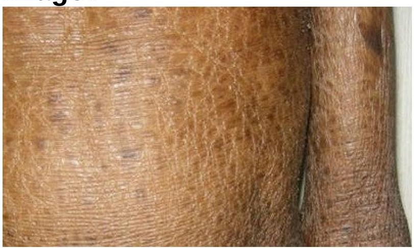
Identify the skin condition depicted in the image.

Which of the following statements is not correct regarding sebaceous cyst?
A cosmetic dermatologist plans to introduce microneedling radiofrequency for acne scars. Which parameter combination would provide optimal collagen remodeling with minimal risk of thermal injury in Fitzpatrick type IV skin?
A 50-year-old man with Fitzpatrick skin type V desires treatment for melasma. He was previously treated with triple combination cream with partial response. What would be the most evidence-based next step considering safety and efficacy?
A patient treated with Q-switched Nd:YAG laser for nevus of Ota develops paradoxical darkening after 4 weeks. What is the most likely explanation for this phenomenon?
A 42-year-old woman develops sudden onset vision loss in one eye 2 hours after hyaluronic acid filler injection in the glabella. Fundoscopy shows retinal whitening. What is the underlying pathophysiology?
Explanation: ***Lichen planus*** - Chemical peels are generally **contraindicated** in active inflammatory conditions like **lichen planus**, as they can worsen the inflammation or trigger a Koebner phenomenon. - While chemical peels can address post-inflammatory hyperpigmentation, they should not be used during the active phase of lichen planus due to the risk of exacerbation. *Melasma* - **Melasma** is a common indication for chemical peels, particularly superficial and medium-depth peels, to reduce hyperpigmentation. - Peels containing agents like **glycolic acid**, salicylic acid, or trichloroacetic acid are often used to lighten melanin deposits. *Acne vulgaris* - Chemical peels are effective in treating **acne vulgaris** by exfoliating the skin, reducing comedones, and improving overall skin texture. - **Salicylic acid peels** are particularly useful due to their lipophilic nature, allowing them to penetrate and clean pores. *Photoaging* - **Photoaging**, characterized by fine lines, wrinkles, and dyspigmentation from sun exposure, is a primary indication for chemical peels. - Peels can promote **collagen remodeling** and help achieve a more even skin tone and smoother texture.
Explanation: ***Actinic keratosis*** - This diagnosis aligns with the description of a **red, scaly plaque** with a **rough texture** and **irregular margins**, which are classic clinical features of actinic keratosis. - The biopsy findings of epidermal and dermal cells with **large, pleomorphic, hyperchromatic nuclei** are consistent with **atypical keratinocytes**, a hallmark of actinic keratosis, indicating **premalignant change**. *Dermal nevus* - A dermal nevus is a **benign melanocytic lesion** that typically presents as a smooth, flesh-colored to light brown papule or nodule, not a scaly or rough plaque. - Histologically, it would show nests of nevus cells primarily in the **dermis** without the significant cellular atypia described. *Junctional nevus* - A junctional nevus is a **benign melanocytic lesion** characterized by nests of nevus cells located at the **dermoepidermal junction**. - Clinically, it appears as a flat or slightly raised, well-demarcated macule or papule, usually uniform in color, lacking the scaly, rough, and irregular features of the presented lesion. *Compound nevus* - A compound nevus is a **benign melanocytic lesion** with nevus cell nests present at both the **dermoepidermal junction** and within the dermis. - It typically presents as a raised, pigmented papule or nodule with a smooth or slightly warty surface, not a scaly plaque with irregular margins.
Explanation: ***Used in photochemotherapy*** - **Methoxysalen** is a **psoralen derivative** that becomes activated by ultraviolet A (UVA) light. - This activation allows it to form **photoadducts with DNA**, inhibiting cell proliferation, which is the basis for its use in **photochemotherapy** for conditions like psoriasis and vitiligo. *Photoprotective agent* - **Photoprotective agents** like sunscreens work by **reflecting or absorbing UV radiation** to prevent skin damage. - **Methoxysalen** actually **sensitizes the skin to UV light**, increasing its effects rather than blocking them. *Melanising agent* - While methoxysalen can induce repigmentation in conditions like vitiligo, it does so by increasing the skin's sensitivity to UV light, which then stimulates **melanogenesis**. - It is not a direct melanising agent that independently promotes melanin production without UV exposure. *Depigmenting agent* - **Depigmenting agents** aim to reduce or remove melanin from the skin, often used for hyperpigmentation disorders. - **Methoxysalen**, especially when used in PUVA therapy, helps to **re-pigment** areas affected by conditions like vitiligo, making it the opposite of a depigmenting agent.
Explanation: ***Discoid lupus erythematosus*** - This condition is a **chronic cutaneous form of lupus** characterized by distinctive **disc-shaped lesions**, often on sun-exposed areas. - **Photosensitivity** is a prominent feature, meaning sunlight direct exposure often **exacerbates or triggers these lesions**. *Chloasma* - This refers to **melasma**, a common condition causing **dark, discolored patches** on the skin, often triggered by **hormonal changes** (e.g., pregnancy) and sun exposure. It does not typically form disc-shaped lesions. - While sunlight exposure influences its presentation, it lacks the characteristic **inflammatory disc-shaped lesions** of discoid lupus. *Dermatitis herpetiformis* - This is an **autoimmune blistering skin condition** strongly associated with **celiac disease**, characterized by intensely pruritic papules and vesicles, not disc-shaped lesions. - Its lesions are **itchy, erythematous papules and vesicles** that are symmetrically distributed, and it is not directly precipitated by sunlight exposure. *Lupus vulgaris* - This is a form of **cutaneous tuberculosis** presenting as slowly progressive, ulcerative, and destructive skin lesions. It is caused by **Mycobacterium tuberculosis** and is unrelated to sun exposure. - It involves direct **tuberculous infection of the skin**, and its clinical presentation differs significantly from the autoimmune, photosensitivity-driven lesions of discoid lupus erythematosus.
Explanation: ***Ichthyosis*** - The image clearly displays widespread **dry, scaling, and thickened skin**, consistent with the characteristic presentation of ichthyosis. - This condition is characterized by a defect in **skin barrier function** leading to excessive dryness and accumulation of scales. *Syndromic ichthyosis* - While syndromic ichthyosis also involves skin scaling, it is associated with **additional systemic symptoms** or **organ involvement**, which cannot be determined from this image alone. - The term "ichthyosis" broadly covers this appearance, and without more clinical information, specifying it as syndromic is not the most direct identification. *Leprosy* - Leprosy typically presents with **hypopigmented, anesthetic skin patches** or **nodules**, which are not seen in the image. - The texture and color changes in the image are not characteristic of the primarily neurological and dermatological manifestations of leprosy. *Cutaneous sarcoidosis* - Cutaneous sarcoidosis manifests as **reddish-brown papules, plaques, or nodules**, often on the face, neck, or extremities. - The widespread, fine scaling and dryness seen in the image do not align with the typical granulomatous lesions of sarcoidosis.
Explanation: ***Treatment is incision and drainage*** - The standard treatment for a sebaceous cyst (more accurately an **epidermoid cyst** or **pilar cyst**) is **surgical excision** of the entire cyst wall to prevent recurrence. - **Incision and drainage** only provides temporary relief by emptying the contents but leaves the cyst wall intact, leading to a high chance of the cyst refilling. *Found on hairy areas of the body* - This statement is generally correct as sebaceous cysts often arise from hair follicles and are common in **hair-bearing areas** like the scalp, face, neck, and trunk. - They occur due to the accumulation of **sebum** and keratin within a blocked or damaged sebaceous gland or hair follicle. *Not found on palms and soles* - This statement is correct because **palms and soles** generally **lack sebaceous glands** and hair follicles, hence sebaceous cysts are typically not found in these locations. - Cysts found in these areas are more likely to be **ganglion cysts** or other types of epidermal inclusion cysts. *It has a punctum* - This statement is often correct; many sebaceous cysts (especially epidermoid cysts) have a visible **central punctum** which represents the occluded pore from which the cyst originated. - This punctum is a **key diagnostic feature** and can sometimes exude a cheesy, foul-smelling material.
Explanation: ***Needle depth 1.5-2 mm, temperature 60-65°C, pulse duration 100-200 ms*** - Optimal **collagen remodeling** occurs when the tissue is heated to **60-65°C**, which triggers the denaturation of proteins and the subsequent production of new collagen and elastin. - A depth of **1.5-2 mm** specifically targets the **papillary and mid-reticular dermis**, while the shorter pulse duration minimizes **Post-Inflammatory Hyperpigmentation (PIH)** in **Fitzpatrick type IV** skin. *Needle depth 3.5 mm, temperature 70°C, pulse duration 1000 ms* - Temperatures reaching **70°C** and very high pulse durations significantly increase the risk of **thermal necrosis** and bulk heating injuries. - A depth of **3.5 mm** is often too deep for standard facial acne scarring and may damage underlying **subcutaneous structures** or cause permanent scarring. *Needle depth 4 mm, temperature 75°C, pulse duration 500 ms* - High temperatures of **75°C** cause excessive tissue coagulation, which can lead to localized **skin burns** and prolonged downtime. - Excessive needle depth combined with high energy delivery poses a severe risk for **atrophic scarring** and pigmentary changes in darker skin types. *Needle depth 0.5 mm, temperature 55°C, pulse duration 50 ms* - A depth of **0.5 mm** is generally insufficient to reach the collagen-rich dermis required for significant improvement of **depressed acne scars**. - A temperature of **55°C** is below the threshold for effective **collagen denaturation**, resulting in suboptimal clinical outcomes for scar revision.
Explanation: ***Q-switched Nd:YAG laser 1064 nm with low fluence*** - This approach, often called **laser toning**, uses a long wavelength that spares the epidermis, making it the safest laser option for **Fitzpatrick skin type V** to avoid **post-inflammatory hyperpigmentation (PIH)**. - It is a clinically sound next step for **recalcitrant melasma** that has only partially responded to first-line therapies like **triple combination cream**. *Fractional CO2 laser resurfacing* - This is an **ablative** treatment that causes significant thermal damage, which carries an unacceptably high risk of **PIH** and scarring in darker skin types. - While effective for skin remodeling, it is generally contraindicated for treating melasma in **type V skin** due to the likelihood of worsening the pigmentation. *Intense pulsed light therapy* - **IPL** uses a broad spectrum of light which is poorly targeted for melasma in dark-skinned individuals and is frequently associated with **rebound hyperpigmentation**. - The melanin in the surrounding **darker skin (Type V)** competes for the energy, leading to a high risk of **thermal burns** and uneven results. *TCA 35% chemical peel* - A 35% concentration of **Trichloroacetic acid (TCA)** is considered a **medium-depth peel**, which is generally too aggressive for patients with Fitzpatrick skin type V. - Medium-depth peels in dark skin types are likely to cause **persistent dyschromia** or permanent **hypopigmentation**, whereas superficial peels (like glycolic or salicylic acid) are safer.
Explanation: ***Increased melanogenesis due to suboptimal fluence*** - Paradoxical darkening in **nevus of Ota** during **Q-switched Nd:YAG** therapy often results from **suboptimal fluence**, which triggers reactive **melanogenesis** instead of destroying the target cells. - This occurs when the energy delivered is sufficient to stimulate **dermal melanocytes** but remains below the threshold required for **selective photothermolysis** and cell destruction. *Delayed clearance in deeper dermal melanocytes* - Delayed clearance typically results in a slow resolution of the lesion rather than an actual **increase in pigmentation** or darkening. - The darkening suggests an active production of **melanin** rather than a passive failure of the lymphatic system to clear debris. *Post-inflammatory hyperpigmentation due to epidermal injury* - **Post-inflammatory hyperpigmentation (PIH)** usually presents as a more generalized or superficial brownish tan following **epidermal damage**. - While common in darker skin types, the term "paradoxical darkening" in the context of dermal lesions specifically refers to the reactive stimulation of **dermal melanocytes**. *Conversion to melanoma* - There is no clinical or histopathological evidence that **Q-switched lasers** induce **malignant transformation** or conversion of a benign nevus to **melanoma**. - While **nevus of Ota** has a small baseline risk of ocular or CNS melanoma, laser-induced darkening is a transient physiological response, not a neoplastic change.
Explanation: ***Retrograde embolization via angular artery to ophthalmic artery*** - Glabellar filler injection can inadvertently enter the **angular artery**, where high injection pressure forces the filler **retrograde** into the **ophthalmic artery**. - Once pressure is released, the filler travels antegrade into the **central retinal artery**, causing occlusion and classic **retinal whitening** due to ischemia. *Compression of supraorbital nerve* - This would lead to **sensory changes** or pain in the forehead region rather than sudden, painless vision loss. - Nerve compression does not explain the **fundoscopic finding** of retinal whitening or vascular compromise. *Direct traumatic optic nerve injury* - The **optic nerve** is located deep within the orbit and is not typically reachable by standard aesthetic needles used in the glabella. - Traumatic injury would likely present with an **afferent pupillary defect** without the characteristic **ischemic retinal whitening** associated with artery occlusion. *Allergic reaction causing optic neuritis* - **Optic neuritis** presents with painful eye movements and inflammatory changes, rather than the hyper-acute vision loss seen in arterial embolization. - A localized allergic reaction to **hyaluronic acid** would cause significant swelling and redness at the injection site rather than sudden **retinal ischemia**.
Get full access to all questions, explanations, and performance tracking.
Start For Free