
Basic Dermatology — MCQs

On this page
Darier's disease is associated with which of the following conditions?
Which pigment, when yellow, brown, or black, shows fluorescence under UV light?
Which one of the following is not a lichenoid reaction?
Regarding Erythema multiforme, all are true except?
Clutch claw and ball appearance is seen in which of the following conditions?
What is the classical presentation described?
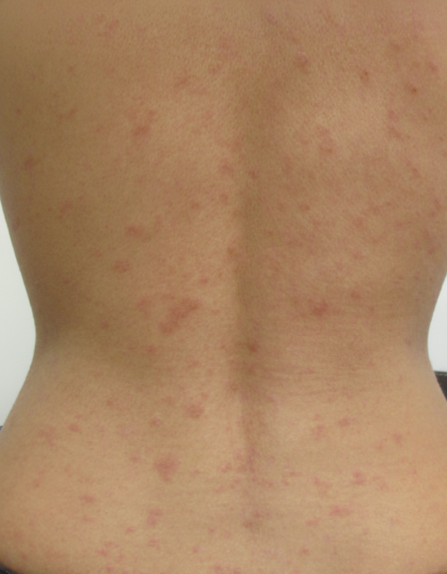
A 50-year-old woman develops pink macules and papules on her hands and forearms in association with a sore throat. The lesions are targetlike, with the centers a dusky violet. A diagnosis of erythema multiforme is made. What is the most important information to obtain from this patient's history?
The Wood's lamp filter is made of?
A Tzanck smear is useful for the diagnosis of which of the following conditions?
Nikolsky's sign is positive in each of the following conditions, EXCEPT:
Practice by Chapter
Structure and Function of Skin
Practice Questions
Cutaneous Histopathology
Practice Questions
Dermatological Examination
Practice Questions
Skin Lesions: Morphology and Description
Practice Questions
Principles of Diagnosis in Dermatology
Practice Questions
Dermatological Procedures
Practice Questions
Wound Healing
Practice Questions
Cutaneous Immunology
Practice Questions
Genetics in Dermatology
Practice Questions
Cutaneous Manifestations of Systemic Diseases
Practice Questions
Geriatric Dermatology
Practice Questions
Pediatric Dermatology Basics
Practice Questions
Want unlimited practice?
Get full access to all questions, explanations, and performance tracking.
Scan to download app