Harlequin ichthyosis is caused by mutation of which gene?
What is the typical duration for pityriasis rosea to resolve?
Where does nevus simplex commonly present?
Sebaceous cysts can occur in all the following locations in the body except:
1 to 2 mm haemorrhages in skin are known as:
Which glands are primarily affected by Fox Fordyce Disease?
Which skin condition is characterized by a 'Christmas tree' appearance?
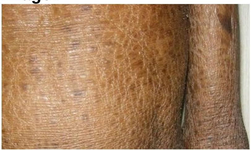
Identify the skin condition depicted in the image.

Which of the following statements is true regarding Acanthosis Nigricans?
Cutis marmorata occurs due to exposure to –
Explanation: ***ABCA12*** - **Harlequin ichthyosis** is a severe, life-threatening genetic skin disorder caused by mutations in the **ABCA12 gene**. - The **ABCA12 gene** encodes an ATP-binding cassette (ABC) transporter protein essential for lipid transport in keratinocytes, critical for epidermal barrier formation. - Mutations lead to defective lamellar granule lipid secretion, resulting in thick, plate-like scales and severe skin abnormalities present at birth. *FAD* - **FAD (Flavin Adenine Dinucleotide)** is a coenzyme, not a gene, making this an inappropriate option. - While FAD is involved in various metabolic redox reactions, it has no direct association with genetic causes of ichthyosis. *Keratin 1* - Mutations in **Keratin 1 (KRT1)** cause **epidermolytic hyperkeratosis** (epidermolytic ichthyosis), characterized by blistering and hyperkeratosis. - This is a different keratinization disorder with a distinct clinical presentation from harlequin ichthyosis. *ALOXE3* - Mutations in **ALOXE3** (arachidonate lipoxygenase 3) cause **autosomal recessive congenital ichthyosis (ARCI)**, also known as **lamellar ichthyosis**. - This presents with collodion membrane at birth and generalized scaling, but is less severe than harlequin ichthyosis.
Explanation: ***6-12 weeks*** - **Pityriasis rosea** is a self-limiting skin condition that typically resolves spontaneously within **6-8 weeks**, though it may take up to 12 weeks in some cases. - The disease progression involves a **herald patch** (present in 50-90% of cases) followed by a generalized secondary eruption that gradually fades over this timeframe. - Most authoritative dermatology texts cite this as the **standard duration** for complete resolution. *1-2 weeks* - This duration is **far too short** for pityriasis rosea, as the secondary rash typically takes 1-2 weeks just to fully develop after the herald patch. - Complete resolution in this timeframe would be extremely unusual and should prompt consideration of alternative diagnoses. *2-4 weeks* - This timeframe is still **too short** for typical resolution, as most patients are still in the active phase of the eruption at 4 weeks. - This might represent the time from herald patch to peak of eruption, not complete resolution. *4-8 weeks* - While some mild cases may begin to resolve around 6-8 weeks, this range is generally considered the **minimum duration** rather than the typical course. - Most standard dermatology references cite 6-8 weeks to 12 weeks as the expected timeframe for complete resolution.
Explanation: ***Face*** - Nevus simplex, also known as a **stork bite**, **angel's kiss**, or **salmon patch**, commonly presents on the face, particularly on the eyelids, glabella, and nuchal area. - These are **capillary malformations** that are present at birth and often fade within the first few years of life. *Trunk* - While other birthmarks can appear on the trunk, nevus simplex has a very strong predilection for the **head and neck region**. - Vascular lesions on the trunk are more likely to be other types, such as infantile hemangiomas, which have a different clinical course. *Legs* - Nevus simplex is **rarely found on the legs**. - Birthmarks on the legs would more typically suggest a different etiology or pattern of vascular malformation. *Hands* - Nevus simplex is **uncommon on the hands**. - Other types of vascular lesions or dermatological conditions might affect the hands but not typically a nevus simplex.
Explanation: ***Palms and soles*** - **Sebaceous cysts** (more accurately termed epidermoid cysts) arise from the **epidermis and hair follicle infundibulum**, not from sebaceous glands themselves. - The **palms and soles** lack both sebaceous glands and hair follicles, making these locations impossible for sebaceous cysts to form. - This is a key anatomical distinction used in clinical diagnosis. *Axilla* - The **axilla** contains numerous hair follicles and sebaceous glands, making it a common site for sebaceous cyst formation. - These cysts can become inflamed or infected due to the moist environment, friction, and bacterial flora of the region. - Hormonal influences can also contribute to cyst development in this area. *Back* - The **back** is a very common location for sebaceous cysts due to a high concentration of hair follicles and sebaceous glands. - Cysts in this area can become quite large and are often subject to irritation from clothing. - The back (especially upper back) is one of the most frequent sites for these lesions. *Pubic area* - The **pubic area** is rich in hair follicles and sebaceous glands, making it susceptible to the formation of sebaceous cysts. - **Hormonal activity** and friction can contribute to the development of these cysts in this region. - Inflammation and infection are more common in this area due to occlusion and moisture.
Explanation: ***Petechiae*** - **Petechiae** are small, pinpoint (<3 mm) hemorrhages into the skin or mucous membranes. - They arise from the leakage of blood from small capillaries, often due to **platelet disorders** or **capillary fragility**. *Micro bleed* - A **micro bleed** typically refers to a small area of bleeding within the brain, detected by neuroimaging, not a skin hemorrhage. - The term is used in the context of brain imaging findings, often associated with **cerebral amyloid angiopathy** or hypertension. *Purpura* - **Purpura** are larger areas of hemorrhage into the skin, typically ranging from 3 mm to 1 cm. - They are caused by similar mechanisms as petechiae but involve a larger volume of extravasated blood, often coalescing to form visible patches. *None of the options* - This option is incorrect because **Petechiae** accurately describes 1-2 mm hemorrhages in the skin.
Explanation: ***Apocrine glands*** - Fox-Fordyce disease is also known as **apocrine miliaria**, directly implicating **apocrine glands** in its pathology. - The disease involves the occlusion of **apocrine duct openings**, leading to rupture of the duct and accumulation of secretory products, causing inflammation and characteristic pruritic papules. *Sebaceous glands* - These glands secrete **sebum** and are primarily affected in conditions like **acne vulgaris** or **sebaceous cysts**, not Fox-Fordyce disease. - While sebaceous glands are present in areas where Fox-Fordyce disease can occur, they are not the primary target of the disease process. *Eccrine Glands* - **Eccrine glands** produce **sweat** for thermoregulation and are affected in conditions like **miliaria crystallina** or **hyperhidrosis**. - Their involvement in Fox-Fordyce disease is secondary or negligible, as the primary pathology is distinct to apocrine structures. *Any gland* - Fox-Fordyce disease specifically targets **apocrine glands**, making a generalized involvement of "any gland" incorrect. - It does not show a predilection for **eccrine** or **sebaceous glands**; the pathology is precise.
Explanation: ***Pityriasis rosea*** - This condition classically presents with an eruption of oval, erythematous, slightly scaly papules and plaques arranged in a **'Christmas tree' pattern** along the skin cleavage lines on the trunk. - It often begins with a single larger lesion known as a **'herald patch'** before the generalized eruption. *Pityriasis rubra pilaris* - Characterized by reddish-orange scaling plaques with **follicular papules** and often involves confluence, leading to areas of normal skin called **'islands of sparing.'** - It does not typically form a 'Christmas tree' pattern; instead, it tends to spread cranially from the lower body. *Psoriasis* - Psoriasis typically presents as well-demarcated, erythematous plaques covered with **silvery scales**, often found on extensor surfaces, scalp, and nails. - The distinctive pattern of psoriasis does not involve a 'Christmas tree' distribution; it's more often characterized by symmetrically distributed lesions. *Vitiligo* - Vitiligo is a pigmentary disorder characterized by **depigmented macules and patches** of varying sizes, resulting from the destruction of melanocytes. - It does not involve inflammatory lesions or any specific 'Christmas tree' arrangement.
Explanation: ***Ichthyosis*** - The image clearly displays widespread **dry, scaling, and thickened skin**, consistent with the characteristic presentation of ichthyosis. - This condition is characterized by a defect in **skin barrier function** leading to excessive dryness and accumulation of scales. *Syndromic ichthyosis* - While syndromic ichthyosis also involves skin scaling, it is associated with **additional systemic symptoms** or **organ involvement**, which cannot be determined from this image alone. - The term "ichthyosis" broadly covers this appearance, and without more clinical information, specifying it as syndromic is not the most direct identification. *Leprosy* - Leprosy typically presents with **hypopigmented, anesthetic skin patches** or **nodules**, which are not seen in the image. - The texture and color changes in the image are not characteristic of the primarily neurological and dermatological manifestations of leprosy. *Cutaneous sarcoidosis* - Cutaneous sarcoidosis manifests as **reddish-brown papules, plaques, or nodules**, often on the face, neck, or extremities. - The widespread, fine scaling and dryness seen in the image do not align with the typical granulomatous lesions of sarcoidosis.
Explanation: ***Is associated with insulin resistant diabetes mellitus.*** - **Acanthosis nigricans** is a common cutaneous marker of **insulin resistance**, frequently seen in patients with **type 2 diabetes mellitus** or prediabetes. - The increased insulin levels stimulate **keratinocyte** and **fibroblast growth factors**, leading to the characteristic skin changes. *Is characterized by hypopigmentation.* - Acanthosis nigricans is characterized by **hyperpigmentation**, presenting as dark, velvety patches, not hypopigmentation. - The darkening of the skin is due to increased melanin production and epidermal thickening. *Commonly occurs in individuals who are lean and thin.* - While it can occur in lean individuals, acanthosis nigricans is **most commonly associated with obesity** and being overweight. - The strong link to **insulin resistance** means it is more prevalent in individuals with higher body mass indices. *Is not typically associated with skin malignancy.* - This statement is **incorrect** - acanthosis nigricans **can be associated with malignancy** as a **paraneoplastic syndrome**, particularly when it has an abrupt onset, rapid progression, or atypical distribution. - It is most commonly associated with **gastrointestinal adenocarcinomas** (e.g., stomach, colon) and sometimes lung or breast cancer.
Explanation: ***Cold temperature*** - **Cutis marmorata** is a physiological response to **cold temperatures**, characterized by a mottled, reticulated vascular pattern on the skin. - This occurs due to **vasoconstriction** of the small arteries and arterioles, alongside **vasodilation** of the venules, creating the characteristic marbled appearance. *Dust* - Exposure to **dust** typically causes **irritation**, allergic reactions, or respiratory issues, such as **dermatitis**, **contact urticaria**, or **asthma**. - It does not directly lead to the characteristic vascular changes seen in cutis marmorata. *Hot temperature* - **Hot temperatures** generally cause **vasodilation** in the skin to facilitate **heat dissipation**, leading to redness and warmth. - This is the opposite physiological response to cutis marmorata, which involves vasoconstriction. *Humidity* - **Humidity** primarily affects **skin hydration** and the rate of perspiration, potentially exacerbating certain skin conditions like **eczema** or **fungal infections**. - High or low humidity does not directly induce the vascular changes that result in cutis marmorata.
Structure and Function of Skin
Practice Questions
Cutaneous Histopathology
Practice Questions
Dermatological Examination
Practice Questions
Skin Lesions: Morphology and Description
Practice Questions
Principles of Diagnosis in Dermatology
Practice Questions
Dermatological Procedures
Practice Questions
Wound Healing
Practice Questions
Cutaneous Immunology
Practice Questions
Genetics in Dermatology
Practice Questions
Cutaneous Manifestations of Systemic Diseases
Practice Questions
Geriatric Dermatology
Practice Questions
Pediatric Dermatology Basics
Practice Questions
Get full access to all questions, explanations, and performance tracking.
Start For Free