A 20-year-old woman presents with the sudden onset of red, target-shaped lesions on her palms and soles, following a herpes simplex outbreak. What is the likely diagnosis?
A 40-year-old man presents with thickened, erythematous skin exhibiting a cobblestone appearance on the lower legs. What is the most likely diagnosis?
A 30-year-old man presents with multiple painful, suppurative nodules in his axillae and groin, and he has a history of smoking. What is the most likely diagnosis?
A 50-year-old male presents with multiple, waxy, brown, stuck-on lesions on his trunk. What is the most likely diagnosis?
Which skin condition is characterized by a 'herald patch'?
Which skin condition is associated with ash leaf spots?
A 30-year-old male presents with recurrent episodes of erythematous target-like lesions, primarily on the hands and forearms, often following herpes simplex outbreaks. What is the most likely diagnosis?
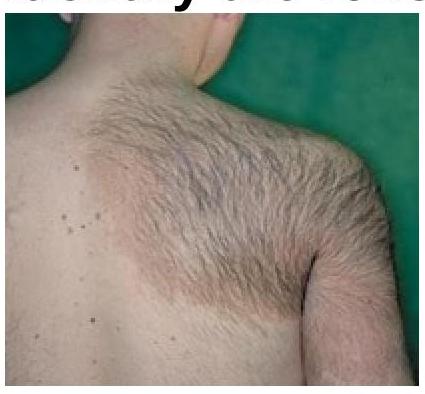
Identify the skin lesion shown in the image.

A known case of diabetes develops annular orange skin lesions that disappear after a biopsy. What is the term used to describe this phenomenon?
Normal epidermal turnover time is?
Explanation: ***Erythema multiforme (EM)*** - This presentation is classic for **Erythema multiforme**, characterized by the acute onset of **target-shaped lesions** on the extremities, often precipitated by **herpes simplex virus (HSV) infection**. - The lesions on the **palms and soles** are a hallmark feature, distinguishing it from other skin conditions. *Stevens-Johnson syndrome (SJS)* - While SJS also involves skin lesions, it is a much more severe condition characterized by **extensive epidermal detachment** (often >10% of body surface area) and significant **mucosal involvement**. - SJS typically involves flu-like prodromal symptoms and is more commonly associated with drug reactions rather than HSV. *Pemphigus vulgaris (PV)* - Pemphigus vulgaris is an **autoimmune blistering disease** causing **flaccid bullae** and erosions on the skin and mucous membranes, often with Nikolsky's sign. - It does not typically present with target lesions nor is it triggered by HSV outbreaks. *Urticaria (hives)* - Urticaria presents as **transient, pruritic, raised wheals** (hives) that can appear anywhere on the body and blanch with pressure. - Unlike erythema multiforme, urticaria lesions are not typically target-shaped and do not commonly persist for days.
Explanation: ***Pretibial myxedema*** * **Pretibial myxedema** presents with **thickened, erythematous skin** on the lower legs, often described as having an "orange peel" or **cobblestone appearance**. * It is specifically associated with **Graves' disease** (hyperthyroidism) due to the deposition of hyaluronic acid and chondroitin sulfate. *Lichen planus* * **Lichen planus** typically manifests as **pruritic, purple, polygonal papules and plaques** (the "5 Ps") rather than thickened, cobblestoned skin. * It commonly affects the wrists, ankles, and oral mucosa, with a different characteristic morphology. *Venous stasis dermatitis* * **Venous stasis dermatitis** also affects the lower legs but presents with **reddish-brown discoloration**, **scaling**, itching, and sometimes ulceration due to chronic venous insufficiency. * The skin changes are more indicative of inflammation and poor circulation, not the distinct thickening or cobblestone texture of myxedema. *Chronic plaque psoriasis* * **Chronic plaque psoriasis** is characterized by well-demarcated, **erythematous plaques** covered with **silvery scales**, primarily affecting extensor surfaces (elbows, knees, scalp). * While it causes thickened skin, the appearance is scaly and not typically described as cobblestone or specifically located only in the pretibial area without other psoriatic manifestations.
Explanation: ***Hidradenitis suppurativa*** - This condition is characterized by **chronic inflammation** of the **apocrine glands**, presenting as painful, deep-seated nodules, abscesses, and sinus tracts in intertriginous areas like the **axillae and groin**. - **Smoking** is a significant risk factor, and the recurring nature of the lesions fits this diagnosis perfectly. *Furunculosis* - **Furunculosis** involves single or multiple painful, pus-filled lesions (boils) caused by bacterial infection of hair follicles, typically **Staphylococcus aureus**. - While furuncles can occur in the axillae and groin, they generally don't present as widespread, chronic, or deeply interconnected **nodules and sinus tracts** characteristic of the patient's presentation. *Lymphogranuloma venereum* - **Lymphogranuloma venereum (LGV)** is a sexually transmitted infection caused by specific serovars of **Chlamydia trachomatis**. - It primarily causes **genital ulcers**, **inguinal lymphadenopathy** (buboes), and proctitis, but does not present with the diffuse, suppurative nodules in the axillae seen in this case. *Erythrasma* - **Erythrasma** is a superficial skin infection caused by **Corynebacterium minutissimum**, characterized by well-demarcated, reddish-brown patches with fine scales, often in intertriginous areas. - It typically causes **asymptomatic or mildly itchy patches** and does not involve painful, suppurative nodules or abscesses.
Explanation: ***Seborrheic keratosis*** - This condition is characterized by **waxy**, **brown**, "stuck-on" lesions, perfectly matching the description. - They are common, **benign epidermal tumors** that typically appear in middle to older age on the trunk, face, or scalp. *Actinic keratosis* - These are **pre-malignant lesions** that are usually rough, scaly patches on sun-exposed skin. - They lack the "stuck-on" appearance and waxy texture described in the patient. *Melanoma* - Melanoma is a **malignant skin cancer** that typically presents as an asymmetrical, irregularly bordered, multi-colored lesion with a changing diameter. - While it can be dark, it usually doesn't have the waxy, "stuck-on" quality of seborrheic keratoses. *Basal cell carcinoma* - This is the most common form of **skin cancer**, often appearing as a pearly nodule with rolled borders, telangiectasias, or a non-healing sore. - It does not present with multiple, waxy, brown, "stuck-on" lesions as described.
Explanation: ***Pityriasis rosea*** - A **herald patch** is the initial, solitary, larger lesion that precedes the generalized rash of pityriasis rosea. - The subsequent rash typically appears in a **Christmas tree pattern** on the trunk. *Psoriasis* - Characterized by well-demarcated, **erythematous plaques** with silvery scales, often on extensor surfaces. - It does not present with a "herald patch"; its onset is typically gradual with the development of multiple lesions. *Lichen planus* - Presents with **pruritic, polygonal, planar, purple papules and plaques** (the 6 P's), often affecting the skin, hair, nails, and mucous membranes. - Does not feature a herald patch as part of its typical presentation. *Eczema* - A broad term for several skin conditions characterized by **itchy, inflamed skin**, often with redness, dryness, and sometimes blistering or weeping. - It does not involve a herald patch; its lesions can be localized or widespread, varying by type (e.g., atopic, contact).
Explanation: ***Tuberous sclerosis*** - **Ash leaf spots** (hypopigmented macules) are a characteristic **cutaneous finding** in tuberous sclerosis, often present from birth. - Tuberous sclerosis is a **neurocutaneous syndrome** also associated with **facial angiofibromas**, **ungual fibromas**, and neurological symptoms like seizures. *Neurofibromatosis* - Characterized by **café-au-lait spots**, which are hyperpigmented macules, not hypopigmented ash leaf spots. - Also associated with **neurofibromas** and **Lisch nodules** (hamartomas of the iris). *Sturge-Weber syndrome* - Primarily presents with a **port-wine stain (facial nevus flammeus)**, which is a vascular malformation, typically unilateral on the face. - Associated with **leptomeningeal angioma** and often causes seizures and glaucoma. *Von Hippel-Lindau disease* - This condition involves the development of **tumors** in various organs, such as **hemangioblastomas** in the brain, spinal cord, and retina, and **renal cell carcinoma**. - It does not characteristically present with skin lesions like ash leaf spots.
Explanation: ***Erythema multiforme*** - Erythema multiforme is characterized by **target-like lesions** with concentric rings of color, typically found on the **hands and forearms**. - It is frequently triggered by **herpes simplex virus (HSV) infection**, which aligns with the recurrent episodes following herpes outbreaks. *Urticaria* - Urticaria (hives) presents as **itchy, transient wheals** that are usually migratory and do not have the classic targetoid appearance. - While it can be recurrent, it is typically an allergic reaction and less commonly directly linked to HSV infections. *Fixed drug eruption* - This condition involves **solitary or a few well-demarcated, often pigmented, erythematous plaques** that recur at the same site(s) with re-exposure to a specific drug. - It does not present with target lesions or a clear association with HSV outbreaks. *Psoriasis* - Psoriasis typically presents with **well-demarcated, erythematous plaques covered by silvery scales**, commonly on extensor surfaces like elbows and knees. - It is a chronic inflammatory condition and does not manifest as acute target lesions or have a direct trigger by HSV outbreaks.
Explanation: ***Becker nevus*** - This image clearly shows a large, **hyperpigmented patch with overlying coarse terminal hairs**, characteristic of a Becker nevus. - Becker nevi typically develop in adolescence and are often found on the shoulder or upper trunk, as seen here. *Hypopigmented macule* - A **hypopigmented macule** would appear as an area of skin with **reduced pigmentation** (lighter than the surrounding skin), which is contrary to the darker lesion shown. - There would also be no indication of **increased hair growth** within a typical hypopigmented macule. *Spitz nevus* - A Spitz nevus is a benign melanocytic nevus often appearing as a **dome-shaped, pink or red papule or nodule**, commonly on the face or limbs. - It does not present as a large, hairy, **hyperpigmented patch** as depicted in the image. *Epidermal nevus* - An epidermal nevus is a **congenital lesion** formed by an overgrowth of epidermal cells, but its appearance is typically a **verrucous (wart-like) plaque** or linearly arranged papules. - While it can be hyperpigmented, it generally **lacks the prominent hypertrichosis** (excessive hair growth) seen in the image.
Explanation: ***Reverse Koebner's phenomenon*** - This phenomenon describes the **improvement or disappearance of skin lesions** at sites of trauma or biopsy, in contrast to the typical Koebner phenomenon where trauma induces new lesions. - In **necrobiosis lipoidica diabeticorum**, a condition associated with diabetes, annular orange lesions may temporarily resolve after trauma, which is characteristic of the reverse Koebner's phenomenon. *Koebner's phenomenon* - Also known as the **isomorphic phenomenon**, it involves the development of **new skin lesions** characteristic of a dermatosis at sites of trauma (e.g., psoriasis, lichen planus, vitiligo). - This is the opposite of what is described in the question, where lesions disappear after biopsy. *Asboe-Hansen sign* - Refers to the **extension of a blister** into previously unaffected skin when pressure is applied to the top of the blister. - This sign is typically observed in **blistering skin conditions** such as pemphigus and bullous pemphigoid, and is unrelated to the resolution of lesions after biopsy. *Isotopic phenomenon* - Describes the manifestation of a **new skin disease** at the site of a prior, unrelated, and already healed skin disease (e.g., herpes zoster followed by lichen planus in the same dermatome). - This phenomenon is not characterized by the disappearance of existing lesions following trauma.
Explanation: ***4 weeks*** - The normal epidermal turnover time, from the basal layer to the shedding of **stratum corneum** cells, is approximately **28 to 30 days**, or roughly 4 weeks. - This duration reflects the time it takes for a keratinocyte to mature and migrate through all layers of the **epidermis**. *1 week* - This period is too short for normal epidermal turnover, as it would suggest a much faster rate of keratinocyte proliferation and differentiation. - A turnover time of 1 week is characteristic of conditions like **psoriasis**, where the epidermal cell cycle is significantly accelerated. *2 weeks* - This timeframe is also shorter than the normal epidermal turnover, implying an accelerated cell cycle, though not as rapid as in conditions like psoriasis. - While cells start migrating earlier, their full maturation and shedding take longer than two weeks. *3 weeks* - While closer to the normal range, 3 weeks still slightly underestimates the full duration of epidermal turnover. - The complete process, including migration through the **stratum granulosum** and formation of a competent stratum corneum, typically extends to 4 weeks.
Structure and Function of Skin
Practice Questions
Cutaneous Histopathology
Practice Questions
Dermatological Examination
Practice Questions
Skin Lesions: Morphology and Description
Practice Questions
Principles of Diagnosis in Dermatology
Practice Questions
Dermatological Procedures
Practice Questions
Wound Healing
Practice Questions
Cutaneous Immunology
Practice Questions
Genetics in Dermatology
Practice Questions
Cutaneous Manifestations of Systemic Diseases
Practice Questions
Geriatric Dermatology
Practice Questions
Pediatric Dermatology Basics
Practice Questions
Get full access to all questions, explanations, and performance tracking.
Start For Free