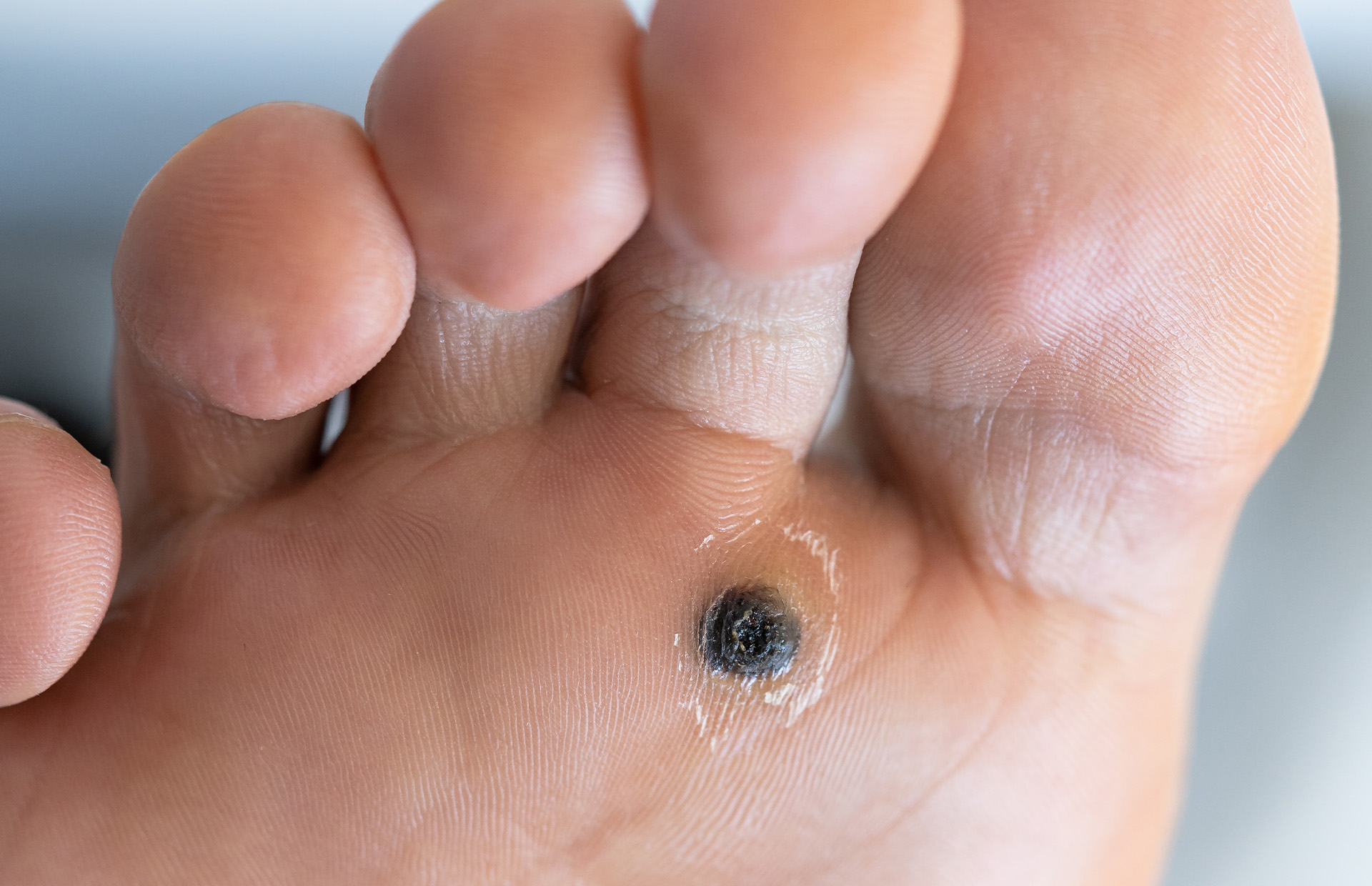
What diagnosis is suggested by the finding on the sole of this patient's foot?

Sawtooth rete ridges are seen in:
Cells as they approach towards stratum corneum show the following features
Koebner's phenomenon seen in ?
Coral red color on Wood's lamp is seen in –
Mucosa is involved in:
Which of the following diseases of the skin is the most likely to be associated with partial anodontia?
Erythema nodosum is seen in all Except
A young boy presented to OPD with multiple shiny pinhead size white papules over dorsum of head, forearm and penis. What would be the diagnosis?
Thickening of the epidermis and/or dermis is
Explanation: ***Plantar wart (Verruca plantaris)*** - The image shows a **hyperkeratotic lesion with central black dots** on the sole of the foot, which is pathognomonic for a **plantar wart** - Plantar warts are caused by **human papillomavirus (HPV)** infection, most commonly types 1, 2, and 4 - The characteristic **black dots represent thrombosed capillaries**, not necrosis - Key diagnostic features include **interruption of skin lines** across the lesion and **tenderness on lateral compression** (pinch test) - They occur on **pressure-bearing areas** of the foot and can be solitary or multiple (mosaic warts) *Chemical burn* - Chemical burns present with **erythema, blistering, or ulceration** with more diffuse or irregular borders - History of **chemical exposure** would be present, and the lesion lacks the characteristic black dots seen in plantar warts - The pattern and location don't fit typical chemical injury *Pemphigus* - Pemphigus is an **autoimmune blistering disease** presenting with **flaccid bullae** that rupture easily - It primarily affects **mucous membranes and flexural areas**, rarely presenting as a solitary hyperkeratotic lesion on the sole - The clinical presentation is completely different from the hyperkeratotic lesion with black dots shown *Radiation dermatitis* - Radiation dermatitis occurs after **therapeutic or accidental radiation exposure** - Presents with **erythema, desquamation, hyperpigmentation, or fibrosis** in the radiation field - Would not present as a discrete hyperkeratotic lesion with black dots, and the sole of the foot is an unusual site for radiation therapy
Explanation: ***Lichen planus*** - **Sawtooth rete ridges** are a characteristic histological feature of **lichen planus**, observed as pointed epidermal rete ridges between which a band-like inflammatory infiltrate is present. - This pattern is due to the destruction of the **basal layer** and subsequent architectural changes in the dermal-epidermal junction. *Psoriasis* - Psoriasis is characterized by **elongated and clubbed rete pegs**, not sawtooth rete ridges. - Other key histological features include **parakeratosis**, **neutrophilic microabscesses**, and **dilated blood vessels** in the dermal papillae. *Erythroderma* - Erythroderma is a clinical presentation of widespread erythema and scaling and not a specific disease with unique histological features. - Histology in erythroderma is **non-specific**, showing features of the underlying causative dermatosis (e.g., psoriasis, eczema) but typically no sawtooth rete ridges on its own. *Darier's disease* - Darier's disease is characterized by **dyskeratosis**, **acantholysis**, and suprabasal clefts. - It presents with distinctive **corps ronds** and **grains** in the epidermis, which are not related to sawtooth rete ridges.
Explanation: ***All of the options*** - As **keratinocytes** differentiate and migrate towards the **stratum corneum**, they undergo a programmed process of maturation and senescence. - This process involves the loss of the nucleus, flattening and dehydration of cells, and significant reduction in mitotic and metabolic activity, all contributing to the protective barrier function of the skin. *Nucleus lost* - The loss of the nucleus is a characteristic feature of **terminal differentiation** of keratinocytes, particularly when they reach the **stratum granulosum** and eventually the stratum corneum. - This anucleated state allows for the formation of a dense, protective layer of **keratin**. *Cells become flat and dehydrated* - As keratinocytes move upwards, they accumulate **keratin filaments** and lose water content, leading to their characteristic flattened and desiccated appearance in the outer layers of the epidermis. - This structural change is crucial for forming a tight, waterproof barrier. *Loss of mitotic and metabolic activity* - Cells in the **stratum corneum** are essentially dead cells, having lost their capacity for division (mitotic activity) and most metabolic processes. - Their primary role shifts from active cellular functions to providing a **physical and chemical barrier**.
Explanation: ***All of the options*** - **Koebner's phenomenon** (isomorphic response) refers to the development of new lesions at sites of **skin trauma** in patients with pre-existing dermatological conditions. - **All four conditions listed** can exhibit Koebner's phenomenon, making this the correct answer. **Psoriasis** - The **most classic and frequently cited** example of Koebner's phenomenon. - Physical injury triggers characteristic red, scaly plaques at trauma sites. - Seen in approximately **25-50%** of psoriasis patients. **Vitiligo** - Well-documented to exhibit **Koebner's phenomenon**. - New **depigmented patches** appear at sites of trauma, cuts, or friction. - Important diagnostic and prognostic indicator in vitiligo patients. **Lichen planus** - Classic condition showing **Koebner's phenomenon**. - New violaceous, flat-topped papules develop at trauma sites. - One of the hallmark features of this condition. **Lichen nitidus** - Although less commonly emphasized, **Lichen nitidus can exhibit Koebner's phenomenon**. - Tiny, shiny papules may appear in linear distribution following trauma. - Part of the lichenoid reaction group that shows isomorphic response.
Explanation: ***Erythrasma*** - **Erythrasma** is a superficial bacterial infection caused by *Corynebacterium minutissimum*, which produces **porphyrins**. - These **porphyrins** fluoresce a characteristic **coral red** under a **Wood's lamp** (UV light of 365 nm). *Vitiligo* - **Vitiligo** causes depigmentation of the skin. Under a Wood's lamp, affected areas appear as a **bright, stark white** or bluish-white due to the complete absence of melanin. - There is no **coral red fluorescence** associated with vitiligo. *Pseudomonas* - Infections caused by **Pseudomonas** (e.g., *Pseudomonas aeruginosa*) can produce pigments that fluoresce under a Wood's lamp, typically appearing **green** or **blue-green**. - This is distinct from the **coral red** seen in Erythrasma. *Tinea* - **Tinea** (dermatophyte) infections may or may not fluoresce under a Wood's lamp; when they do, species like *Microsporum canis* or *Microsporum audouinii* typically produce a **blue-green** or **apple-green** fluorescence. - Most superficial dermatophyte infections, such as those caused by *Trichophyton* species, **do not fluoresce** at all.
Explanation: ***Lichen planus*** - **Lichen planus** frequently involves the mucous membranes, especially the **oral mucosa**, presenting as **Wickham's striae** (lacy white patterns). - Genital, esophageal, laryngeal, and conjunctival mucosa can also be affected, leading to erosions or papules. - This is the **classic dermatological condition** with significant mucosal involvement. *Scabies* - **Scabies** is a parasitic skin infestation caused by the **mite** *Sarcoptes scabiei*, primarily affecting the skin, especially intertriginous areas. - It does not involve mucous membranes, as the mites burrow into the epidermis only. *Psoriasis* - **Psoriasis** is predominantly a **skin disorder** characterized by red, scaly plaques. - Mucosal involvement is **extremely rare** (unlike lichen planus where it is common). - While geographic tongue and rare oral lesions have been described, typical mucosal involvement is not a feature of psoriasis. *Alopecia* - **Alopecia** refers to **hair loss** and is a condition primarily affecting hair follicles in the scalp or other hairy areas of the body. - It does not involve the mucous membranes, which are non-hairy epithelial linings.
Explanation: ***Hereditary ectodermal dysplasia*** - This condition is characterized by abnormal development of **ectodermal structures**, including teeth, hair, nails, and sweat glands. - **Partial anodontia** (the congenital absence of some teeth) or even complete anodontia (**oligodontia**) is a hallmark clinical feature of ectodermal dysplasia due to impaired tooth bud formation. *Erythema multiforme* - This is an **acute, self-limiting inflammatory reaction** of the skin and mucous membranes, often triggered by infections (e.g., HSV) or medications. - It presents with characteristic **target lesions** and mucosal erosions but is not associated with dental developmental abnormalities like anodontia. *Lichen planus* - This is a **chronic inflammatory mucocutaneous disorder** affecting the skin, hair, nails, and mucous membranes, often presenting with **pruritic, purple, polygonal papules**. - It is an immune-mediated condition and does not involve developmental defects of teeth. *Keratosis follicularis* - Also known as **Darier disease**, this is an **autosomal dominant disorder** characterized by abnormal keratinization, leading to greasy, crusted papules primarily in seborrheic areas. - While it affects skin appendages, it is not directly linked to tooth agenesis or partial anodontia.
Explanation: ***Salicylate poisoning*** - **Salicylate poisoning** is a toxic condition resulting from an overdose of aspirin or related compounds and is not associated with erythema nodosum. - Symptoms primarily involve **gastrointestinal disturbance**, **tinnitus**, **metabolic acidosis**, and **respiratory alkalosis**. *Tuberculosis* - **Erythema nodosum** can be a manifestation of primary **tuberculosis**, representing a hypersensitivity reaction to mycobacterial antigens. - It often occurs in children or young adults with recent **Mycobacterium tuberculosis** infection. *Leprosy* - **Erythema nodosum leprosum** is a specific type of erythema nodosum seen in **leprosy**, particularly during type 2 lepra reactions. - It presents as multiple, tender, erythematous nodules often accompanied by **fever** and **systemic symptoms**. *Typhoid* - **Typhoid fever** (enteric fever) can rarely be associated with **erythema nodosum** as a cutaneous manifestation of the systemic infection caused by **Salmonella Typhi**. - Other skin findings in typhoid include **rose spots**, which are more characteristic.
Explanation: ***Lichen nitidus*** - Characterized by **multiple, shiny, pinpoint, flesh-colored to whitish papules**, often seen on the upper extremities, trunk, genitalia, and flexural areas. - The lesions are typically **asymptomatic** but can sometimes be pruritic. *Scabies* - Presents with intensely **pruritic** papules, vesicles, and burrows, especially in interdigital spaces, wrists, and waistline. - The lesions are usually *erythematous* and excoriated due to scratching, which does not match the description of shiny white papules. *Lichen planus* - Typically presents as **pruritic, purple, polygonal, planar papules and plaques** (the "6 Ps"). - Often associated with **Wickham's striae** (fine white lines) and can affect mucous membranes, nails, and hair, which is distinct from the described lesions. *Molluscum contagiosum* - Characterized by **dome-shaped, flesh-colored, pearly papules** with a central **umbilication** caused by a poxvirus. - The lesions are usually larger than "pinhead size" and have a characteristic central dimple not described here.
Explanation: ***Lichenification*** - **Lichenification** is the correct answer as it involves thickening of **both the epidermis (acanthosis)** and **dermis (fibrosis)**, which directly matches the question asking for thickening of "epidermis and/or dermis" - Characterized by leathery thickening with exaggerated skin markings - Results from chronic scratching or rubbing leading to epidermal hyperplasia and dermal fibrosis *Hyperkeratosis* - **Hyperkeratosis** refers only to thickening of the **stratum corneum** (the outermost keratinized layer), not the entire epidermis or dermis - It is limited to increased keratin in the cornified layer - Does not involve the full thickness of epidermis or any dermal thickening *Callus* - A **callus** is a localized form of hyperkeratosis affecting only the stratum corneum - Forms in response to repeated friction or pressure - Represents localized thickening, not generalized epidermal or dermal thickening *Scaling* - **Scaling** refers to visible shedding or flaking of dead skin cells from the stratum corneum - It is a descriptive term for desquamation, not thickening - Does not indicate epidermal or dermal thickening
Structure and Function of Skin
Practice Questions
Cutaneous Histopathology
Practice Questions
Dermatological Examination
Practice Questions
Skin Lesions: Morphology and Description
Practice Questions
Principles of Diagnosis in Dermatology
Practice Questions
Dermatological Procedures
Practice Questions
Wound Healing
Practice Questions
Cutaneous Immunology
Practice Questions
Genetics in Dermatology
Practice Questions
Cutaneous Manifestations of Systemic Diseases
Practice Questions
Geriatric Dermatology
Practice Questions
Pediatric Dermatology Basics
Practice Questions
Get full access to all questions, explanations, and performance tracking.
Start For Free