
Basic Dermatology — MCQs

On this page
Patients with which of the following conditions are at greatest risk of pernio
Sebaceous cysts can occur in all the following locations in the body except:
What is the color of tuberous sclerosis lesions when examined under a Wood's lamp?
Keratodermic sandals are associated with which of the following conditions?
Which skin condition is characterized by a 'Christmas tree' appearance?
Which glands are primarily affected by Fox Fordyce Disease?
Which of the following statements is true regarding Acanthosis Nigricans?
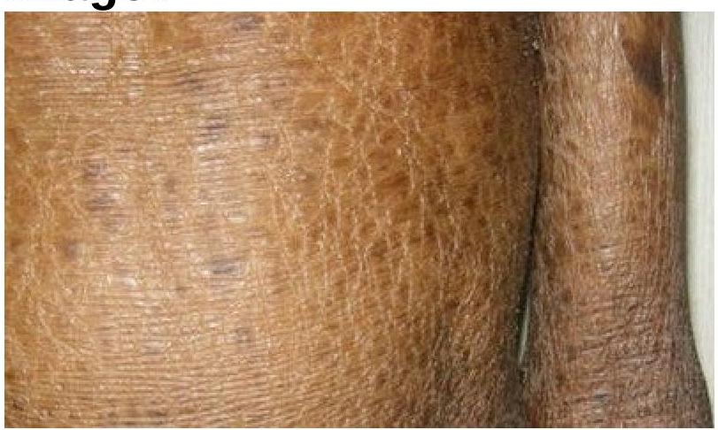
Identify the skin condition depicted in the image.
Cutis marmorata occurs due to exposure to –
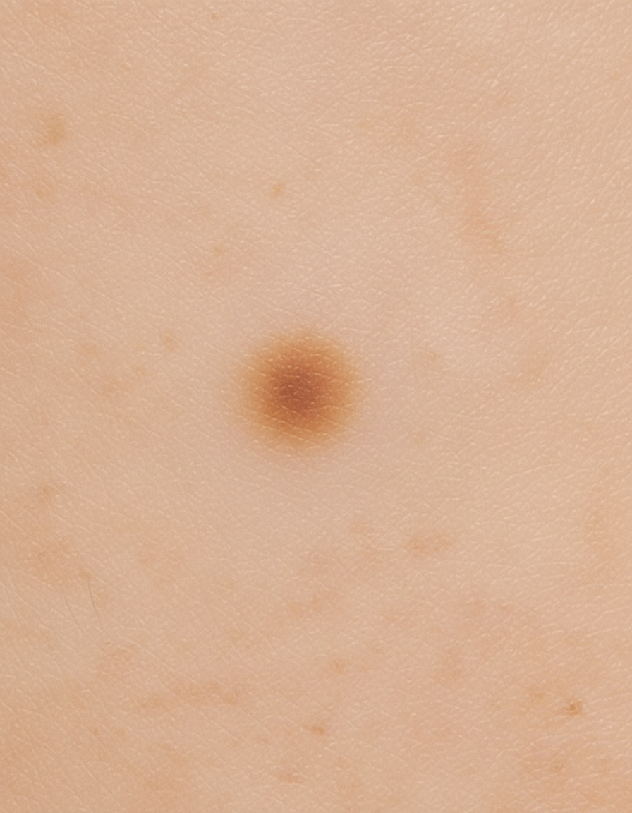
Identify the type of skin lesion shown in the image.
Practice by Chapter
Structure and Function of Skin
Practice Questions
Cutaneous Histopathology
Practice Questions
Dermatological Examination
Practice Questions
Skin Lesions: Morphology and Description
Practice Questions
Principles of Diagnosis in Dermatology
Practice Questions
Dermatological Procedures
Practice Questions
Wound Healing
Practice Questions
Cutaneous Immunology
Practice Questions
Genetics in Dermatology
Practice Questions
Cutaneous Manifestations of Systemic Diseases
Practice Questions
Geriatric Dermatology
Practice Questions
Pediatric Dermatology Basics
Practice Questions
Want unlimited practice?
Get full access to all questions, explanations, and performance tracking.
Scan to download app