
Basic Dermatology — MCQs

On this page
A 35-year-old female with a history of chronic cough and dyspnea presents with multiple erythematous nodules on her shins. What is the most likely diagnosis?
A 45-year-old man presents with multiple firm, yellowish nodules on his extensor surfaces. What is the most likely diagnosis?
Which skin condition is characterized by a 'herald patch'?
A 30-year-old male presents with recurrent episodes of erythematous target-like lesions, primarily on the hands and forearms, often following herpes simplex outbreaks. What is the most likely diagnosis?
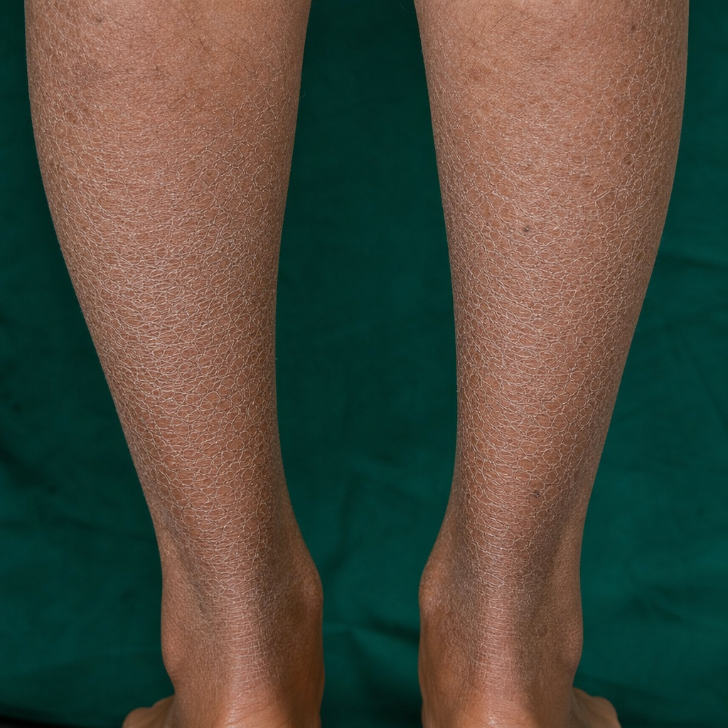
Identify the skin condition shown in the image.
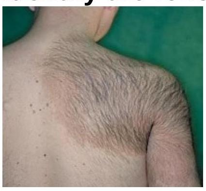
Identify the skin lesion shown in the image.
Normal epidermal turnover time is?
A known case of diabetes develops annular orange skin lesions that disappear after a biopsy. What is the term used to describe this phenomenon?
What is the typical duration for pityriasis rosea to resolve?
Harlequin ichthyosis is caused by mutation of which gene?
Practice by Chapter
Structure and Function of Skin
Practice Questions
Cutaneous Histopathology
Practice Questions
Dermatological Examination
Practice Questions
Skin Lesions: Morphology and Description
Practice Questions
Principles of Diagnosis in Dermatology
Practice Questions
Dermatological Procedures
Practice Questions
Wound Healing
Practice Questions
Cutaneous Immunology
Practice Questions
Genetics in Dermatology
Practice Questions
Cutaneous Manifestations of Systemic Diseases
Practice Questions
Geriatric Dermatology
Practice Questions
Pediatric Dermatology Basics
Practice Questions
Want unlimited practice?
Get full access to all questions, explanations, and performance tracking.
Scan to download app