
Basic Dermatology — MCQs

On this page
Epidermal turnover time in healthy adults is
Thickening of the epidermis and/or dermis is
All of the following are not true with respect to erythema multiforme except?
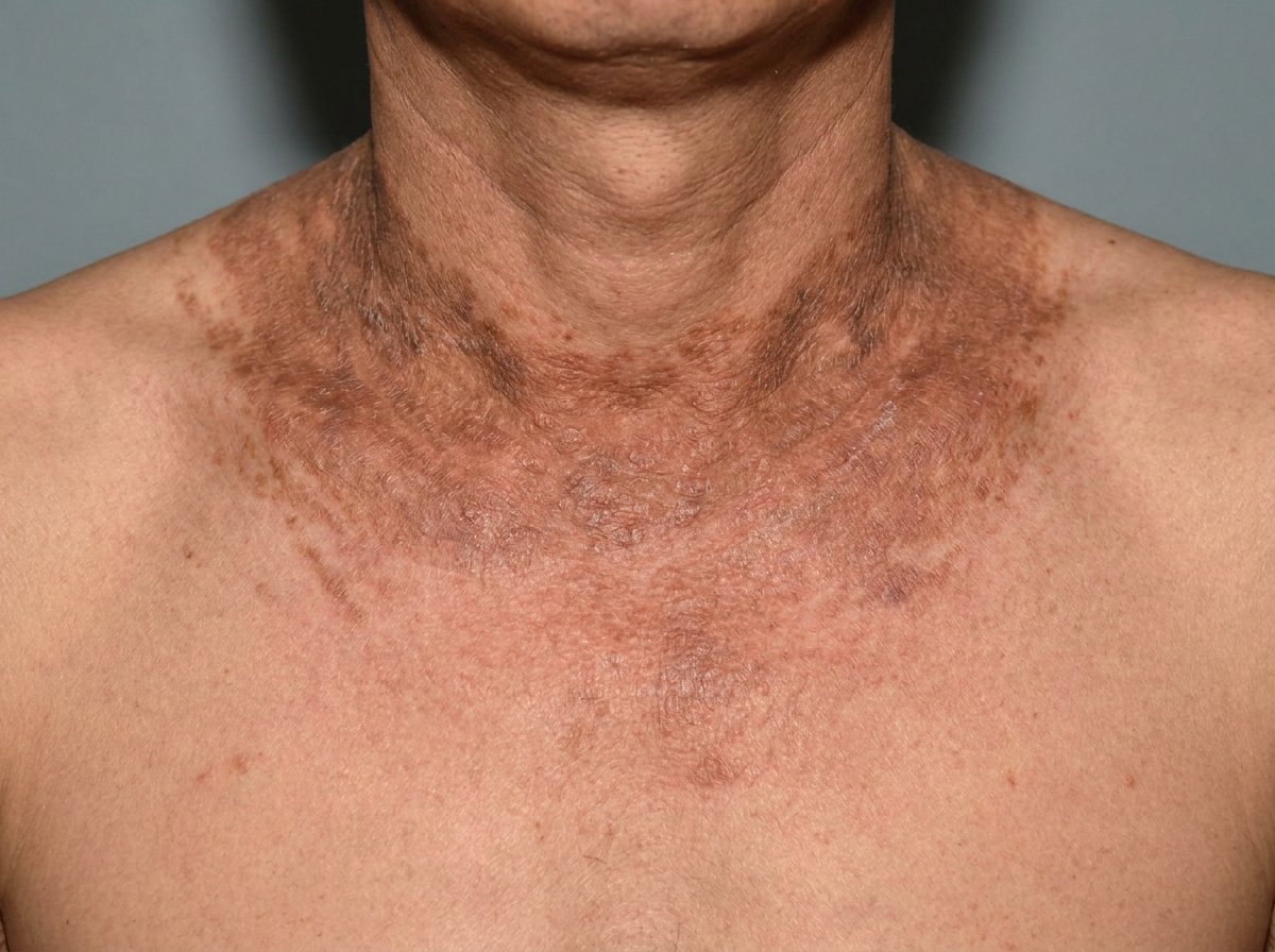
Dermatological manifestation of which of the following diseases?
Which is TRUE about dermatologic emergencies?
A 35-year-old female presents with recurrent painful nodules on shins, associated with fever and arthralgia. Previous biopsy showed septal panniculitis. She's now pregnant at 20 weeks. Most appropriate treatment is:
Patient on anti-TB drugs develops tender nodules on shins. Most likely diagnosis is:
All are true about lichen planus EXCEPT:
Which of the following is a typical feature of erythema multiforme?
A 20-year-old woman presents with the sudden onset of red, target-shaped lesions on her palms and soles, following a herpes simplex outbreak. What is the likely diagnosis?
Practice by Chapter
Structure and Function of Skin
Practice Questions
Cutaneous Histopathology
Practice Questions
Dermatological Examination
Practice Questions
Skin Lesions: Morphology and Description
Practice Questions
Principles of Diagnosis in Dermatology
Practice Questions
Dermatological Procedures
Practice Questions
Wound Healing
Practice Questions
Cutaneous Immunology
Practice Questions
Genetics in Dermatology
Practice Questions
Cutaneous Manifestations of Systemic Diseases
Practice Questions
Geriatric Dermatology
Practice Questions
Pediatric Dermatology Basics
Practice Questions
Want unlimited practice?
Get full access to all questions, explanations, and performance tracking.
Scan to download app