
Basic Dermatology — MCQs

On this page
Which of the following is NOT true about angioneurotic edema?
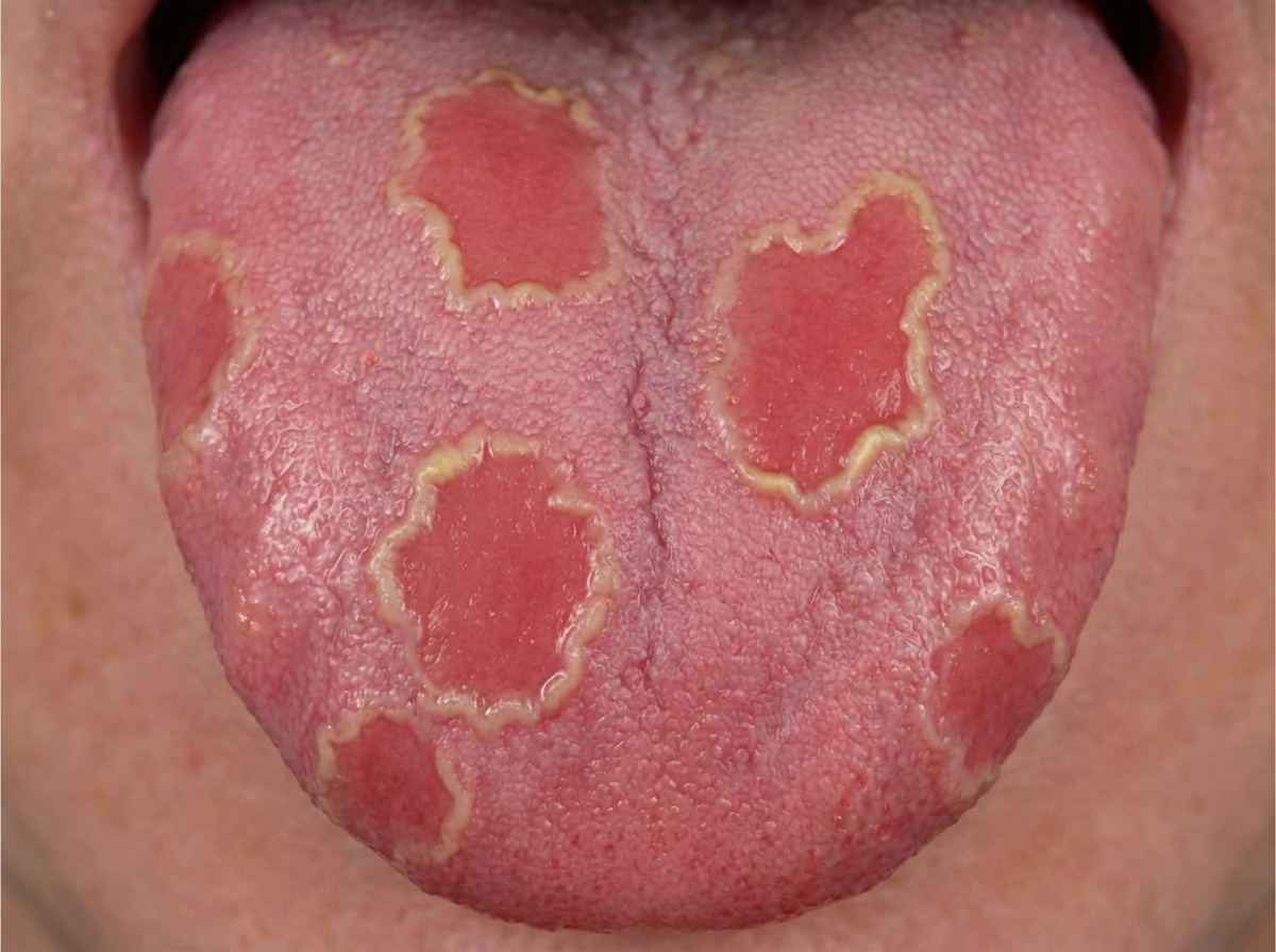
A patient with a history of smoking, essential hypertension, hypercholesterolemia, and hyperuricemia presents for follow-up. Oral examination findings from three visits are available. What is the most likely diagnosis of the tongue involvement?
A 40-year-old diabetic patient presents with round lesions on the abdomen for 2 weeks. Histopathology shows a palisading granuloma. What is the diagnosis?
Leg ulcers are typically associated with which of the following conditions, EXCEPT?
Palpable purpura is seen in which of the following conditions?
What is the wavelength of light emitted by a Wood's lamp?
A child presents with recurrent mouth ulcers that typically begin as a round, yellowish, elevated spot surrounded by a red halo and heal within 7-10 days. What is the most likely diagnosis?
All of the following conditions are a differential diagnosis for Koebner Phenomenon, EXCEPT:
An 85-year-old male cigar smoker with no notable medical history presented with black discoloration and hairy appearance of the tongue, which had lasted several years. He denied using bismuth-containing compounds. What is the cause of this lesion on the tongue?
A 24-year-old male presents with a skin condition. What is the provisional diagnosis for this condition?

Practice by Chapter
Structure and Function of Skin
Practice Questions
Cutaneous Histopathology
Practice Questions
Dermatological Examination
Practice Questions
Skin Lesions: Morphology and Description
Practice Questions
Principles of Diagnosis in Dermatology
Practice Questions
Dermatological Procedures
Practice Questions
Wound Healing
Practice Questions
Cutaneous Immunology
Practice Questions
Genetics in Dermatology
Practice Questions
Cutaneous Manifestations of Systemic Diseases
Practice Questions
Geriatric Dermatology
Practice Questions
Pediatric Dermatology Basics
Practice Questions
Want unlimited practice?
Get full access to all questions, explanations, and performance tracking.
Scan to download app