
Basic Dermatology — MCQs

On this page
A 22-year-old woman presents with multiple tender, erythematous nodules on her shins that developed over the past week. She reports having a sore throat 2 weeks ago. She also complains of joint pain and fatigue. Physical examination reveals raised, red, tender nodules on the anterior surface of both legs. Her temperature is 38.2°C. Which of the following is the most likely diagnosis?
A 28-year-old Caucasian woman presents to a local walk-in clinic with the complaint of pruritus and a salmon-colored scaling patch on her back. The patient stated that she developed a cold a couple of weeks ago and that her skin lesion has enlarged in the last week. The past medical history is unremarkable. The physical examination reveals a generalized exanthem, bilateral symmetric macules pointing towards the cleavage lines, and a salmon-colored patch on her back, with a well-demarcated border containing a collarette with fine-scale. What is the best next step of management in this case?
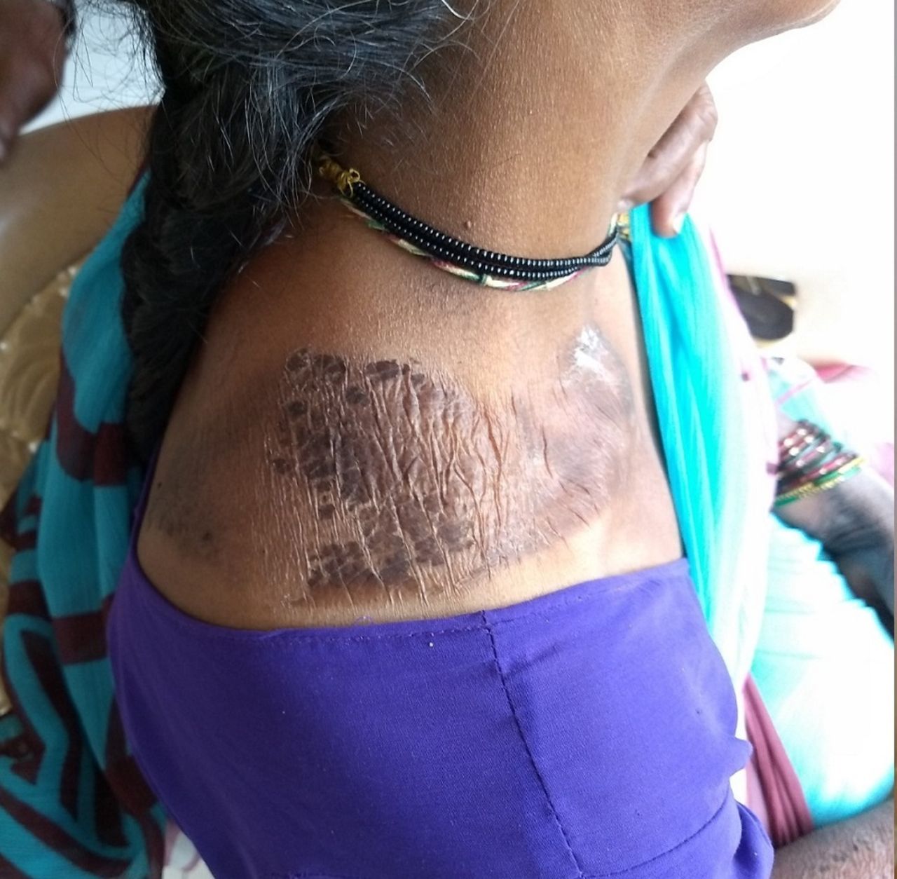
What is the name of this appearance which is seen in pellagra?
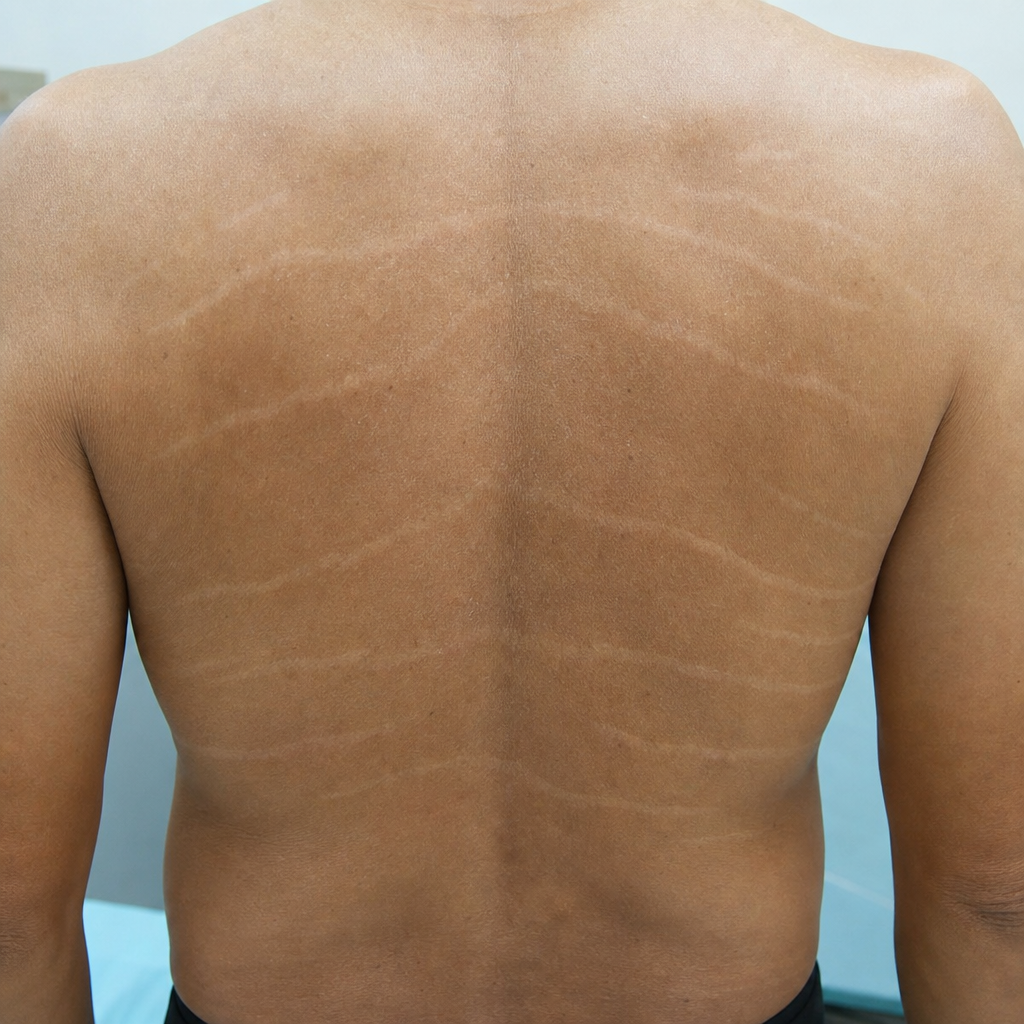
What are these horizontal lines seen on the skin called?
Wood's lamp is not used for diagnosing:
Granular layer is absent in:
Diascopy is very helpful in the diagnosis of:
Which is a secondary skin lesion?
Perleche is caused by:
Keratodermic sandals is a feature of ?
Practice by Chapter
Structure and Function of Skin
Practice Questions
Cutaneous Histopathology
Practice Questions
Dermatological Examination
Practice Questions
Skin Lesions: Morphology and Description
Practice Questions
Principles of Diagnosis in Dermatology
Practice Questions
Dermatological Procedures
Practice Questions
Wound Healing
Practice Questions
Cutaneous Immunology
Practice Questions
Genetics in Dermatology
Practice Questions
Cutaneous Manifestations of Systemic Diseases
Practice Questions
Geriatric Dermatology
Practice Questions
Pediatric Dermatology Basics
Practice Questions
Want unlimited practice?
Get full access to all questions, explanations, and performance tracking.
Scan to download app