
Basic Dermatology — MCQs

On this page
Pruritus is not seen in which of the following conditions?
The condition shown in the color plate can be seen in which of the following syndromes?
A young male presented with scaly truncal lesions. On examination, genital lesions along with oral 'lace-like' lesions are seen. What is the most likely diagnosis?
A 38-year-old woman presented with a 2-week history of tender, swollen nodules on her lower legs. Her only current medication was a triphasic oral contraceptive. She denies any history of inflammatory bowel disease, sarcoidosis, or chronic infections. Physical examination revealed erythematous nodular swellings on the anterior lower legs. A punch biopsy specimen obtained from a lesion on the left anterior tibia reveals a septal inflammation of subcutaneous fat. What is the best description of this rash?
Thin zone of Grenz is seen in which of the following conditions?
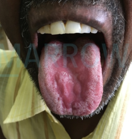
A young lady presents with white lacy lesions in the oral cavity and her proximal nail fold has extended onto the nail bed. What is the likely diagnosis?
What is the most common malignancy associated with ichthyosis?
A 16-year-old female presents with a one-week history of small ulcers on her cheek and tongue, accompanied by a mild fever. Oral examination reveals multiple ulcers on the buccal and labial mucosa. She reports that her board examinations are in one week. What is the most appropriate management for this patient?
All are true about the features of erythromelalgia, EXCEPT:
A 40-year-old woman presents with a burning sensation in the mouth. Clinical examination reveals lesions consisting of radiating white striations in a retiform arrangement affecting the buccal mucosa, tongue, lips, and gingiva bilaterally. An incisional biopsy is suggestive of lichen planus. Which of the following is NOT a clinical form of Lichen Planus?
Practice by Chapter
Structure and Function of Skin
Practice Questions
Cutaneous Histopathology
Practice Questions
Dermatological Examination
Practice Questions
Skin Lesions: Morphology and Description
Practice Questions
Principles of Diagnosis in Dermatology
Practice Questions
Dermatological Procedures
Practice Questions
Wound Healing
Practice Questions
Cutaneous Immunology
Practice Questions
Genetics in Dermatology
Practice Questions
Cutaneous Manifestations of Systemic Diseases
Practice Questions
Geriatric Dermatology
Practice Questions
Pediatric Dermatology Basics
Practice Questions
Want unlimited practice?
Get full access to all questions, explanations, and performance tracking.
Scan to download app