
Basic Dermatology — MCQs

On this page
Keloid formation is not typically seen over which of the following areas?
The pustular, neutrophil-rich skin lesions that develop or worsen following intradermal trauma is called what?
Oral examination of a 57-year-old female reveals a white patch on the buccal mucosa that cannot be scraped off. She has no features suggestive of immunosuppression. What is the most likely diagnosis?
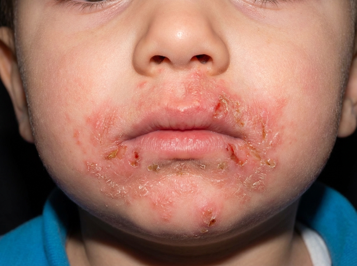
The given condition is caused due to deficiency of which of the following?
Saw tooth rete pegs are seen in which of the following conditions?
Angioid streaks in the eyes are seen in which of the following conditions?
Which of the following is NOT a component of Naxos syndrome?
A 12-year-old girl presents for a routine physical examination. The patient has numerous freckles over her upper trunk and face. Which of the following terms best describes the morphologic appearance of her freckles?
A 70-year-old male presented with an asymptomatic white patch on the oral cavity following the application of a denture. What is the treatment of choice?
Which of the following conditions is due to a defect in the normal keratinization of the oral mucosa?
Practice by Chapter
Structure and Function of Skin
Practice Questions
Cutaneous Histopathology
Practice Questions
Dermatological Examination
Practice Questions
Skin Lesions: Morphology and Description
Practice Questions
Principles of Diagnosis in Dermatology
Practice Questions
Dermatological Procedures
Practice Questions
Wound Healing
Practice Questions
Cutaneous Immunology
Practice Questions
Genetics in Dermatology
Practice Questions
Cutaneous Manifestations of Systemic Diseases
Practice Questions
Geriatric Dermatology
Practice Questions
Pediatric Dermatology Basics
Practice Questions
Want unlimited practice?
Get full access to all questions, explanations, and performance tracking.
Scan to download app