
Basic Dermatology — MCQs

On this page
What is the initial treatment of a keloid?
All of the following are true regarding erythrocyanosis frigida, EXCEPT:
What is the primary function of Langerhans cells?
Which of the following conditions does not show an isomorphic type of Koebner phenomenon?
A young lady presents with lacy lesions in the oral cavity and genitals, and her proximal nail fold has extended onto the nail bed. What is the likely diagnosis?
What are the characteristic bodies found in Lichen planus?
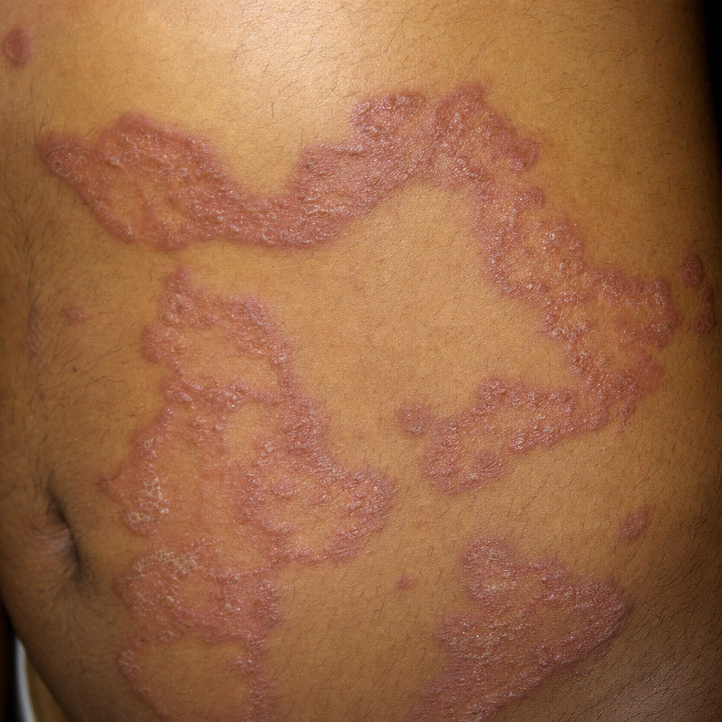
A patient with a 4-year history of diabetes presents with the skin lesion shown in the image. What is the most likely diagnosis?
Migratory necrolytic erythema is seen in which of the following conditions?
What is the recommended treatment for geographic tongue?
All of the following perforating disorders are associated with chronic renal failure EXCEPT:
Practice by Chapter
Structure and Function of Skin
Practice Questions
Cutaneous Histopathology
Practice Questions
Dermatological Examination
Practice Questions
Skin Lesions: Morphology and Description
Practice Questions
Principles of Diagnosis in Dermatology
Practice Questions
Dermatological Procedures
Practice Questions
Wound Healing
Practice Questions
Cutaneous Immunology
Practice Questions
Genetics in Dermatology
Practice Questions
Cutaneous Manifestations of Systemic Diseases
Practice Questions
Geriatric Dermatology
Practice Questions
Pediatric Dermatology Basics
Practice Questions
Want unlimited practice?
Get full access to all questions, explanations, and performance tracking.
Scan to download app