Identify the skin lesion shown in the image.

Diascopy is very helpful in the diagnosis of:
A child has a rash. His family history is positive for asthma. What could be the most probable diagnosis?

A 30-year-old male presented with silvery scales on elbow and knee, that bleed on removal. The probable diagnosis is:
Koebner's phenomenon seen in ?
Which of the following methods is used for demonstrating old washed bloodstains?
A farmer presented with a black mole on the cheek. It increased in size, more than 6mm with irregular borders and a central black lesion, what could be the diagnosis?
Dyskeratosis refers to which of the following?
What is the optimal wavelength of light emitted by a Wood's lamp for dermatological examinations?
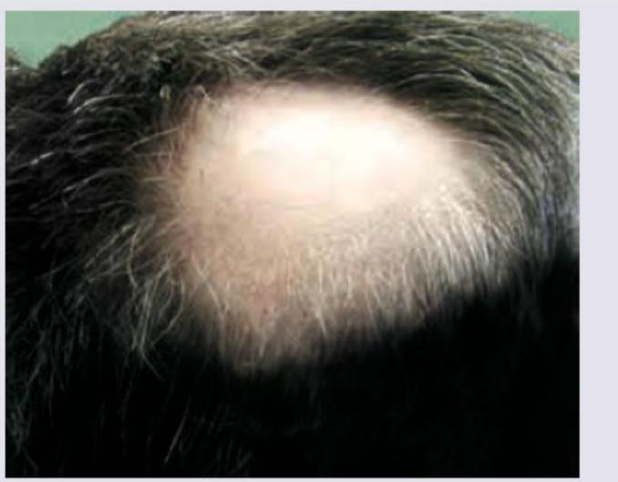
In a patient with the following lesion on scalp, what changes are seen in the nails?

Explanation: ***Becker nevus*** - This image clearly shows a large, **hyperpigmented patch with overlying coarse terminal hairs**, characteristic of a Becker nevus. - Becker nevi typically develop in adolescence and are often found on the shoulder or upper trunk, as seen here. *Hypopigmented macule* - A **hypopigmented macule** would appear as an area of skin with **reduced pigmentation** (lighter than the surrounding skin), which is contrary to the darker lesion shown. - There would also be no indication of **increased hair growth** within a typical hypopigmented macule. *Spitz nevus* - A Spitz nevus is a benign melanocytic nevus often appearing as a **dome-shaped, pink or red papule or nodule**, commonly on the face or limbs. - It does not present as a large, hairy, **hyperpigmented patch** as depicted in the image. *Epidermal nevus* - An epidermal nevus is a **congenital lesion** formed by an overgrowth of epidermal cells, but its appearance is typically a **verrucous (wart-like) plaque** or linearly arranged papules. - While it can be hyperpigmented, it generally **lacks the prominent hypertrichosis** (excessive hair growth) seen in the image.
Explanation: ***All of the options*** - Diascopy is a diagnostic technique using a glass slide to apply pressure on skin lesions, helping differentiate between **vascular (erythematous)** and **non-vascular lesions** and revealing underlying pathology. - It is particularly helpful in diagnosing **lupus vulgaris**, **cutaneous vasculitis**, and **nevus anaemicus**. *Lupus vulgaris* - Shows pathognomonic **apple-jelly nodules** on diascopy due to granulomatous inflammation. - When pressed with a glass slide, the lesion reveals a characteristic **translucent yellowish-brown ("apple jelly") color** from tuberculoid granulomas. *Cutaneous vasculitis* - Diascopy differentiates **purpura (extravasated blood)** from simple erythema. - **Non-blanching purpura** indicates intravascular hemorrhage from vessel wall damage, a key feature of vasculitis. - Blanching erythema would suggest vasodilation rather than true vasculitis. *Nevus anaemicus* - Diascopy causes the pale lesion to **disappear or blend** with surrounding blanched normal skin. - This occurs because normal surrounding vessels constrict under pressure, matching the baseline pale appearance of the nevus. - This helps distinguish it from other hypopigmented lesions like vitiligo (which remains visible on diascopy).
Explanation: ***Atopic dermatitis*** - The presence of a rash in a child with a family history of **asthma** strongly suggests atopic dermatitis, as it is part of the **atopic triad** (eczema, asthma, allergic rhinitis). - Atopic dermatitis often presents with **erythematous, pruritic patches** and plaques, commonly affecting flexural areas like the antecubital and popliteal fossae, as well as the face and neck in younger children. *Seborrheic dermatitis* - This condition typically presents with **greasy, yellowish scales** on an erythematous base, often affecting areas rich in sebaceous glands such as the scalp, face (nasolabial folds), and chest. - While it can occur in infants, it does not have the strong association with a family history of asthma seen in atopic dermatitis. *Allergic contact dermatitis* - This rash results from an **exposure to an allergen**, leading to a localized, erythematous, and pruritic eruption, often with vesicles or bullae, at the site of contact. - The history does not provide information about a specific allergen exposure, and while it could produce a similar-looking rash, the family history of asthma points more strongly to atopic diathesis. *Erysipelas* - Erysipelas is a superficial skin infection, usually caused by *Streptococcus pyogenes*, presenting as a **well-demarcated, intensely erythematous, warm, and painful rash** with a raised border. - This is an **acute bacterial infection** and would typically be accompanied by systemic symptoms like fever and chills, which are not mentioned in the child's presentation.
Explanation: ***Psoriasis*** - The presence of **silvery scales** on the elbows and knees, which **bleed upon removal** (Auspitz sign), is a classic presentation of **plaque psoriasis**. - Psoriasis is a chronic inflammatory skin condition characterized by **accelerated epidermal turnover**. *Secondary syphilis* - Secondary syphilis typically presents with a **generalized maculopapular rash**, which can affect the palms and soles, but it does not usually feature silvery scales or the Auspitz sign. - Other common symptoms of secondary syphilis include **fever, lymphadenopathy, and condyloma lata**. *Pityriasis* - **Pityriasis rosea** is characterized by an oval, fawn-colored, scaly rash, often preceded by a **herald patch**, and usually resolves spontaneously. It does not typically present with silvery scales or bleeding on removal. - **Pityriasis versicolor** is caused by yeast and presents as hypopigmented or hyperpigmented macules with fine scales, commonly on the trunk, not silvery scales on elbows and knees. *Seborrhoeic dermatitis* - Seborrhoeic dermatitis involves greasy, yellowish scales on red skin, typically affecting areas rich in sebaceous glands like the scalp, face (nasolabial folds, eyebrows), and chest. - It does not present with silvery scales or the Auspitz sign, which are specific to psoriasis.
Explanation: ***All of the options*** - **Koebner's phenomenon** (isomorphic response) refers to the development of new lesions at sites of **skin trauma** in patients with pre-existing dermatological conditions. - **All four conditions listed** can exhibit Koebner's phenomenon, making this the correct answer. **Psoriasis** - The **most classic and frequently cited** example of Koebner's phenomenon. - Physical injury triggers characteristic red, scaly plaques at trauma sites. - Seen in approximately **25-50%** of psoriasis patients. **Vitiligo** - Well-documented to exhibit **Koebner's phenomenon**. - New **depigmented patches** appear at sites of trauma, cuts, or friction. - Important diagnostic and prognostic indicator in vitiligo patients. **Lichen planus** - Classic condition showing **Koebner's phenomenon**. - New violaceous, flat-topped papules develop at trauma sites. - One of the hallmark features of this condition. **Lichen nitidus** - Although less commonly emphasized, **Lichen nitidus can exhibit Koebner's phenomenon**. - Tiny, shiny papules may appear in linear distribution following trauma. - Part of the lichenoid reaction group that shows isomorphic response.
Explanation: **Luminol spray** - **Luminol** reacts with the iron in **hemoglobin** to produce a blue-white luminescence, making it highly effective for detecting even heavily diluted or rinsed-away bloodstains. - It is particularly useful for demonstrating **old, washed-up bloodstains** at crime scenes where visual identification might be difficult. *Infrared photography* - While useful for detecting certain hidden details or substances, **infrared photography** is not the primary method for revealing old or washed-up bloodstains. - **Bloodstains** can absorb infrared light to varying degrees, but the chemical reaction of luminol is specifically designed for trace blood detection. *Magnifying lens* - A **magnifying lens** merely enhances the visibility of existing stains or patterns and cannot detect traces of blood invisible to the naked eye, particularly old or diluted ones. - It is a tool for closer inspection, not for chemical detection of hidden substances. *Ultraviolet light* - **Ultraviolet (UV) light** can be used to detect certain biological fluids such as semen or saliva, which **fluoresce** under UV. - However, fresh or old bloodstains typically *absorb* UV light rather than fluoresce, making it less effective for detecting them, especially if they are washed up.
Explanation: ***Superficial spreading melanoma*** - This is the most common type of melanoma and often presents as a **mole with irregular borders**, varying colors, and a diameter greater than 6mm, consistent with the description. - The lesion typically grows **radially** across the skin surface before beginning vertical growth, indicated by the increase in size. *Acral lentigo melanoma* - This type of melanoma primarily affects the **palms, soles, and nail beds**, which is inconsistent with a lesion on the cheek. - It often appears as a **dark brown or black patch** that slowly enlarges, but its location is characteristic. *Lentigo maligna melanoma* - This melanoma typically occurs in **chronically sun-damaged skin** of the elderly, often on the head and neck, but usually presents as a **flat, irregularly shaped, tan or brown patch** with varying shades, which may not fit the description of a central black lesion within a larger mole. - It has a dominant **radial growth phase** and progresses slowly over many years before developing a nodular component. *Nodular melanoma* - This type is characterized by its **rapid vertical growth** and appearance as a **raised, dark, often dome-shaped lesion** from the outset. - While it can be black, the description of an "increased in size" mole with irregular borders and a central black lesion points more towards a spreading type rather than a rapidly growing nodule from the beginning.
Explanation: ***Abnormal, premature keratinization within cells below the stratum granulosum.*** - **Dyskeratosis** is a histological term for **premature keratinization** of individual keratinocytes. - This typically occurs in cells that are *below* the **stratum granulosum**, often in the stratum spinosum, indicating abnormal differentiation. *Discontinuity of the skin showing incomplete loss of epidermis.* - This description refers to an **erosion** or **ulceration**, depending on the depth of the epidermal loss. - It does not specifically describe abnormal cellular keratinization. *Keratinization with retained nuclei in the stratum corneum.* - This is the definition of **parakeratosis**, which is a normal finding in mucous membranes but an abnormal finding in skin, often associated with disorders like **psoriasis**. - It indicates incomplete maturation of keratinocytes as they reach the stratum corneum. *Thickening of stratum corneum, often associated with a qualitative abnormality of keratin.* - This describes **hyperkeratosis**, which is an increase in the thickness of the **stratum corneum**. - While it can involve abnormal keratin, it refers to increased thickness, not premature individual cell keratinization.
Explanation: **365 nm** - A Wood's lamp primarily emits **long-wave UVA light** in the 320 to 400 nm range, with an optimal peak around **365 nm**. - This specific wavelength is ideal for inducing **fluorescence** in various dermatological conditions, making them visible. *400 nm* - While within the UVA range, **400 nm** is at the higher end and may not provide the optimal fluorescence yield for all diagnostic purposes compared to 365 nm. - Light at 400 nm is closer to the visible light spectrum and might offer less distinction for subtle fluorescence. *320 nm* - **320 nm** is at the lower end of the UVA spectrum, bordering on UVB. - While still capable of inducing some fluorescence, it is generally less effective than 365 nm for the conditions typically examined with a Wood's lamp. *200 nm* - **200 nm** falls into the **UVC range** (100-280 nm), which is harmful and not used for diagnostic purposes in a Wood's lamp. - This wavelength is absorbed by the atmosphere and epidermis and can cause significant **DNA damage**, making it unsafe for routine dermatological examination.
Explanation: ***Pitting of nails*** - The image shows a patch of **alopecia areata** on the scalp. **Nail pitting** is the most common and characteristic nail change associated with alopecia areata, occurring in **10-66% of cases**. - Pitting appears as small depressions or **"ice-pick" marks** on the nail surface, resulting from defective nail matrix keratinization. - Other nail changes in alopecia areata include **trachyonychia (rough nails), red spotted lunulae, onycholysis**, and **Beau's lines**. *Dorsal pterygium of nails* - **Dorsal pterygium** occurs when the proximal nail fold fuses with and extends over the nail plate, creating a wing-like scar. - This is classically associated with **lichen planus, trauma, burns, vasculitis**, and **graft-versus-host disease** — **NOT alopecia areata**. - It can lead to permanent nail dystrophy or nail loss. *Azure nails* - **Azure nails** (blue nails) are typically associated with **Wilson's disease** (copper accumulation) or **minocycline use**, not alopecia areata. - They represent a blue-gray discoloration of the nail bed or lunula. *Yellow nail discolouration* - **Yellow nail syndrome** is a rare condition characterized by slow-growing, thickened, yellow nails, often associated with **lymphedema** and **respiratory problems** (pleural effusions, chronic bronchitis). - It is not linked to alopecia areata.
Get full access to all questions, explanations, and performance tracking.
Start For Free