What is the primary function of Langerhans' cells?
A 32 year old man presents with a 3-month history of weight loss, night sweats, a productive cough with blood-tinged sputum, anorexia, general malaise, and a low grade fever. A PPD skin test shows > 10 mm of induration. If the area of induration were biopsied, which of the following type of reactive cells would be found?
A 60-year-old female presents with eczematous itching lesions. Biopsy revealed a subepidermal cleft with Direct Immunofluorescence showing Linear C3 & IgG deposition along the basement membrane zone. What is the likely diagnosis?
Statement 1 - A 59-year-old patient presents with flaccid bullae. Histopathology shows a suprabasal acantholytic split. Statement 2 - The row of tombstones appearance is diagnostic of Pemphigus vulgaris.
Most common precipitant of contact dermatitis is?
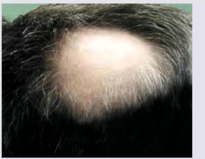
In a patient with the following lesion on scalp, what changes are seen in the nails?

In which of the following conditions is the Koebner phenomenon most commonly observed?
Patient on anti-TB drugs develops tender nodules on shins. Most likely diagnosis is:
Lines of Blaschko are related to?
Which sign is pathognomonic for neurofibromatosis?
Explanation: ***Antigen presenting cells*** - Langerhans' cells play a critical role as **antigen presenting cells** (APCs) in the immune system, facilitating the activation of T-cells [1][2]. - They are found in the **epidermis** and are essential in initiating immune responses against pathogens [1][3]. *Seen in auto immune conditions* - While Langerhans' cells may be involved in autoimmune responses, they are not exclusively seen in these conditions. - Their primary function isn't linked to autoimmunity but rather to **immunological surveillance** and **antigen presentation** [1]. *Phagocytic cells* - Langerhans' cells are not primarily **phagocytic**, as their main role focuses on presenting antigens rather than directly engulfing pathogens [1]. - Phagocytic cells include macrophages and neutrophils, which are more involved in **directly consuming foreign particles**. *Seen in chronic infection* - Although Langerhans' cells can participate in the immune response during infections, they are not specifically characterized as being prominent in **chronic infections**. - Chronic infections are typically associated with different immune cell dynamics, involving other cells such as **plasma cells** and **T-cells**. **References:** [1] Kumar V, Abbas AK, et al.. Robbins and Cotran Pathologic Basis of Disease. 9th ed. Diseases of the Immune System, p. 200. [2] Kumar V, Abbas AK, et al.. Robbins and Cotran Pathologic Basis of Disease. 9th ed. Diseases of the Immune System, pp. 207-208. [3] Kumar V, Abbas AK, et al.. Robbins and Cotran Pathologic Basis of Disease. 9th ed. The Skin, p. 1144.
Explanation: ***T lymphocyte*** - The clinical picture (weight loss, night sweats, productive cough with blood-tinged sputum, positive PPD) is highly suggestive of **tuberculosis**, a **Type IV hypersensitivity reaction** [1], [2]. - **Type IV hypersensitivity reactions** are cell-mediated, involving the activation of **T lymphocytes**, which migrate to the site of antigen exposure (like a PPD test site or a tuberculous granuloma) and release cytokines, leading to induration and inflammation [1], [2]. *Eosinophil* - **Eosinophils** are primarily involved in allergic reactions and defense against parasitic infections [3]. - They are not the predominant reactive cells in a **Type IV hypersensitivity** response like that seen in tuberculosis [1]. *Mast cell* - **Mast cells** play a critical role in immediate hypersensitivity reactions (Type I), releasing histamine and other mediators [4]. - They are not the primary cells involved in the delayed-type hypersensitivity response elicited by tuberculin purified protein derivative (PPD) [2]. *B lymphocyte* - **B lymphocytes** are responsible for humoral immunity by producing antibodies [3]. - While they contribute to overall immune responses, they are not the main effector cells in a cell-mediated **Type IV hypersensitivity reaction** characteristic of a positive PPD test [1], [2]. **References:** [1] Cross SS. Underwood's Pathology: A Clinical Approach. 6th ed. (Basic Pathology) introduces the student to key general principles of pathology, both as a medical science and as a clinical activity with a vital role in patient care. Part 2 (Disease Mechanisms) provides fundamental knowledge about the cellular and molecular processes involved in diseases, providing the rationale for their treatment. Part 3 (Systematic Pathology) deals in detail with specific diseases, with emphasis on the clinically important aspects., pp. 173-174. [2] Kumar V, Abbas AK, et al.. Robbins and Cotran Pathologic Basis of Disease. 9th ed. Diseases of the Immune System, p. 218. [3] Cross SS. Underwood's Pathology: A Clinical Approach. 6th ed. (Basic Pathology) introduces the student to key general principles of pathology, both as a medical science and as a clinical activity with a vital role in patient care. Part 2 (Disease Mechanisms) provides fundamental knowledge about the cellular and molecular processes involved in diseases, providing the rationale for their treatment. Part 3 (Systematic Pathology) deals in detail with specific diseases, with emphasis on the clinically important aspects., pp. 195-196. [4] Kumar V, Abbas AK, et al.. Robbins and Cotran Pathologic Basis of Disease. 9th ed. Diseases of the Immune System, pp. 208-210.
Explanation: ***Bullous Pemphigoid*** - The presence of **eczematous itching lesions**, a **subepidermal cleft**, and **linear C3 and IgG deposition along the basement membrane zone** on direct immunofluorescence (DIF) are classic diagnostic features of Bullous Pemphigoid. - This autoimmune blistering disease typically affects older individuals and is characterized by antibodies targeting components of the **hemidesmosomes**, specifically BP180 and BP230. *Pemphigus foliaceus* - This condition involves **intraepidermal blistering**, specifically within the granular layer, rather than a subepidermal cleft. - DIF in Pemphigus foliaceus shows **intercellular IgG deposition** in the epidermis, not linear deposition along the basement membrane zone. *Pemphigus Vulgaris* - Pemphigus Vulgaris is characterized by **intraepidermal blistering** above the basal cell layer (**suprabasal clefting**), leading to fragile bullae that rupture easily. - DIF typically reveals **intercellular IgG and C3 deposition** in a "chicken wire" pattern throughout the epidermis, which differs from the linear pattern seen in this case. *Dermatitis herpetiformis* - While Dermatitis herpetiformis is also an autoimmune blistering disease with itching lesions, its characteristic DIF finding is **granular IgA deposition** in the dermal papillae, not linear C3 and IgG at the basement membrane zone. - Histopathology in Dermatitis herpetiformis shows **subepidermal vesicles** with neutrophil infiltration in the dermal papillae, but the direct immunofluorescence pattern is distinct.
Explanation: ***Correct: Statements 1 & 2 are correct, 2 is not explaining 1*** **Analysis of Statement 1:** - A 59-year-old patient with **flaccid bullae** and **suprabasal acantholytic split** on histopathology is the classic presentation of **Pemphigus vulgaris** - The flaccid (easily ruptured) nature of bullae distinguishes it from tense bullae seen in bullous pemphigoid - The suprabasal location of the split (just above the basal layer) with acantholysis (loss of cell-to-cell adhesion) is pathognomonic - **Statement 1 is CORRECT** ✓ **Analysis of Statement 2:** - The **"row of tombstones" or "tombstone appearance"** is indeed a diagnostic histopathological feature of Pemphigus vulgaris - This appearance results from basal keratinocytes remaining attached to the basement membrane while suprabasal cells separate due to acantholysis - The intact basal cells standing upright resemble a row of tombstones - **Statement 2 is CORRECT** ✓ **Does Statement 2 explain Statement 1?** - Statement 2 describes a **histopathological appearance** (tombstone pattern) that is a **consequence** of the suprabasal split - However, it does NOT explain the **underlying cause** of the flaccid bullae or the suprabasal split - The true explanation involves **IgG autoantibodies against desmoglein 3 (and desmoglein 1)**, which attack intercellular adhesion structures (desmosomes), causing **acantholysis** - Therefore, **Statement 2 does NOT explain Statement 1** ✗ *Incorrect: Statement 2 is the correct explanation for Statement 1* - While both statements describe features of Pemphigus vulgaris, the tombstone appearance is a descriptive finding, not an explanatory mechanism *Incorrect: Statements 1 and 2 are incorrect* - Both statements are medically accurate descriptions of Pemphigus vulgaris features *Incorrect: Statement 1 is incorrect* - Statement 1 correctly describes the cardinal clinical and histopathological features of Pemphigus vulgaris
Explanation: ***Nickel*** - **Nickel** is the most frequent cause of **allergic contact dermatitis**, commonly found in jewelry, belt buckles, and zippers. - Exposure leads to a **Type IV hypersensitivity reaction**, characterized by erythema, itching, and vesiculation. *Gold* - While gold can cause contact dermatitis, it is **far less common** than nickel allergy. - Reactions to gold are often seen with prolonged skin contact, such as with jewelry. *Silver* - **Silver** is a **rare cause** of allergic contact dermatitis. - Allergic reactions to silver are typically observed in individuals with extensive exposure, such as jewelers. *Iron* - **Iron** is **not a common precipitant** of contact dermatitis. - Allergic reactions to iron are exceedingly rare, as iron is an essential element found naturally in the body.
Explanation: ***Pitting of nails*** - The image shows a patch of **alopecia areata** on the scalp. **Nail pitting** is the most common and characteristic nail change associated with alopecia areata, occurring in **10-66% of cases**. - Pitting appears as small depressions or **"ice-pick" marks** on the nail surface, resulting from defective nail matrix keratinization. - Other nail changes in alopecia areata include **trachyonychia (rough nails), red spotted lunulae, onycholysis**, and **Beau's lines**. *Dorsal pterygium of nails* - **Dorsal pterygium** occurs when the proximal nail fold fuses with and extends over the nail plate, creating a wing-like scar. - This is classically associated with **lichen planus, trauma, burns, vasculitis**, and **graft-versus-host disease** — **NOT alopecia areata**. - It can lead to permanent nail dystrophy or nail loss. *Azure nails* - **Azure nails** (blue nails) are typically associated with **Wilson's disease** (copper accumulation) or **minocycline use**, not alopecia areata. - They represent a blue-gray discoloration of the nail bed or lunula. *Yellow nail discolouration* - **Yellow nail syndrome** is a rare condition characterized by slow-growing, thickened, yellow nails, often associated with **lymphedema** and **respiratory problems** (pleural effusions, chronic bronchitis). - It is not linked to alopecia areata.
Explanation: ***Correct: Psoriasis*** - **Psoriasis** is the **most classic and commonly cited example** of the Koebner phenomenon (isomorphic response) - New psoriatic plaques characteristically develop at sites of cutaneous trauma, scratches, or surgical incisions in 25-50% of psoriasis patients - This is a **pathognomonic feature** frequently tested in competitive exams and considered the prototype condition for demonstrating this phenomenon - The mechanism involves inflammatory cascades triggered by trauma in genetically predisposed skin *Incorrect: Lichen planus* - While lichen planus does exhibit the Koebner phenomenon with purplish polygonal papules appearing along scratch lines, it is **less commonly observed** compared to psoriasis - Seen in approximately 10-25% of lichen planus cases - Not considered the primary example when teaching about Koebner phenomenon *Incorrect: Viral warts* - Viral warts can demonstrate **pseudo-Koebner phenomenon** where new warts form along trauma lines due to viral inoculation - This is more accurately described as **autoinoculation** rather than true isomorphic response - Less commonly discussed in the context of classic Koebner phenomenon compared to psoriasis *Incorrect: All of the options* - While all three conditions can show Koebner-like responses, the question asks for "**most commonly observed**" - Psoriasis remains the **gold standard** and most frequently encountered example in clinical practice and medical literature
Explanation: ***Erythema nodosum*** - Erythema nodosum is a common **cutaneous adverse drug reaction** to anti-TB medications, presenting with **tender, erythematous nodules** typically on the shins. - It is a form of **panniculitis** (inflammation of subcutaneous fat) specifically associated with various triggers, including infections and drugs, making it highly probable in this context. *Sweet syndrome* - Sweet syndrome (acute febrile neutrophilic dermatosis) presents with **tender, erythematous plaques and nodules** often associated with fever and leukocytosis. - While it can be drug-induced, it typically involves a more widespread skin eruption and prominent systemic symptoms like **fever**, which are not specified here. *Panniculitis* - Panniculitis is a general term for **inflammation of the subcutaneous fat**, and erythema nodosum is a type of panniculitis. - This option is too broad; while accurate, "Erythema nodosum" is the **most specific and likely diagnosis** given the patient’s presentation in the context of anti-TB drug use. *Erythema multiforme* - Erythema multiforme is characterized by **target lesions** (concentric rings of erythema and edema) and often involves mucous membranes. - The description of **tender nodules on shins** does not fit the characteristic morphology of erythema multiforme.
Explanation: ***Keratinocytes*** - **Lines of Blaschko** represent the migratory pathways of embryonic cells, primarily **keratinocytes**, in the skin. - These lines are not visible under normal conditions but become apparent in various **genetic skin disorders** where abnormal cells follow these specific patterns. *Blood vessels* - While blood vessels are extensively present in the skin, they do not follow the specific **migratory patterns** described by the Lines of Blaschko. - Their arrangement is more related to **vascular networks** and anatomical supply rather than embryonic cell migration. *Nerves* - **Nerves** in the skin have specific distributions, often following dermatomal patterns, which are distinct from the **Lines of Blaschko**. - Nerve distribution is related to their segmental origin from the **spinal cord**, not the migratory paths of epidermal cells. *Bones* - **Bones** are part of the skeletal system and are not found in the skin, making them unrelated to the **Lines of Blaschko**. - These lines describe epidermal cell migration, which is a feature of the **integumentary system**.
Explanation: **Explanation:** **Neurofibromatosis Type 1 (NF1)**, also known as von Recklinghausen disease, is an autosomal dominant multisystem disorder. The correct answer is **Axillary freckling (Crowe sign)** because it is considered highly specific (pathognomonic) for NF1. 1. **Axillary Freckling (Crowe Sign):** These are small, 1–3 mm hyperpigmented macules found in intertriginous areas (axilla or groin). Unlike solar lentigines, they appear in areas not exposed to the sun. Their presence is a hallmark diagnostic criterion for NF1. 2. **Cafe-au-lait macules (CALMs):** While these are often the first sign of NF1, they are **not pathognomonic**. CALMs can be seen in healthy individuals, McCune-Albright syndrome, Fanconi anemia, and Legius syndrome. In NF1, the presence of 6 or more macules (>5mm in prepubertal; >15mm in postpubertal) is required for diagnosis. 3. **Shagreen patch:** This is a connective tissue nevus (leathery plaque) typically found on the lower back. It is a characteristic feature of **Tuberous Sclerosis**, not Neurofibromatosis. **High-Yield Clinical Pearls for NEET-PG:** * **Lisch Nodules:** Iris hamartomas (seen on slit-lamp exam) are the most common ocular finding in NF1. * **Optic Glioma:** The most common CNS tumor associated with NF1. * **Sphenoid Wing Dysplasia:** A classic skeletal deformity in NF1. * **Genetics:** NF1 is caused by a mutation in the *NF1* gene on **Chromosome 17** (encodes Neurofibromin), while NF2 is linked to **Chromosome 22** (encodes Merlin).
Get full access to all questions, explanations, and performance tracking.
Start For Free