Which of the following is NOT a characteristic of pemphigus vulgaris?
A skin biopsy shows acantholysis with intraepidermal blistering. Which immunofluorescence pattern would confirm pemphigus vulgaris?
A 40 year old male reported with recurrent episodes of oral ulcers, large areas of denuded skin and flaccid vesiculo-bullous eruptions. Which is the most important bedside investigation helpful in establishing the diagnosis -
Which of the following statements about mucous membrane pemphigoid is correct?
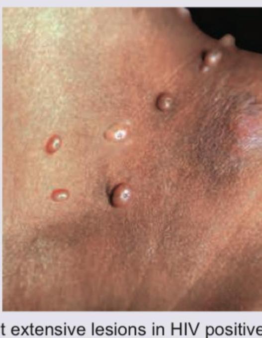
A patient presents with the skin lesions shown in the image. While evaluating for possible blistering disorders, all of the following conditions could present with similar morphology EXCEPT:

A child presents with grouped vesicles on an erythematous base on the buttocks. What is the most likely diagnosis?
A skin biopsy shows suprabasilar acantholysis with 'row of tombstones' appearance. Which immunofluorescence pattern would confirm pemphigus vulgaris?
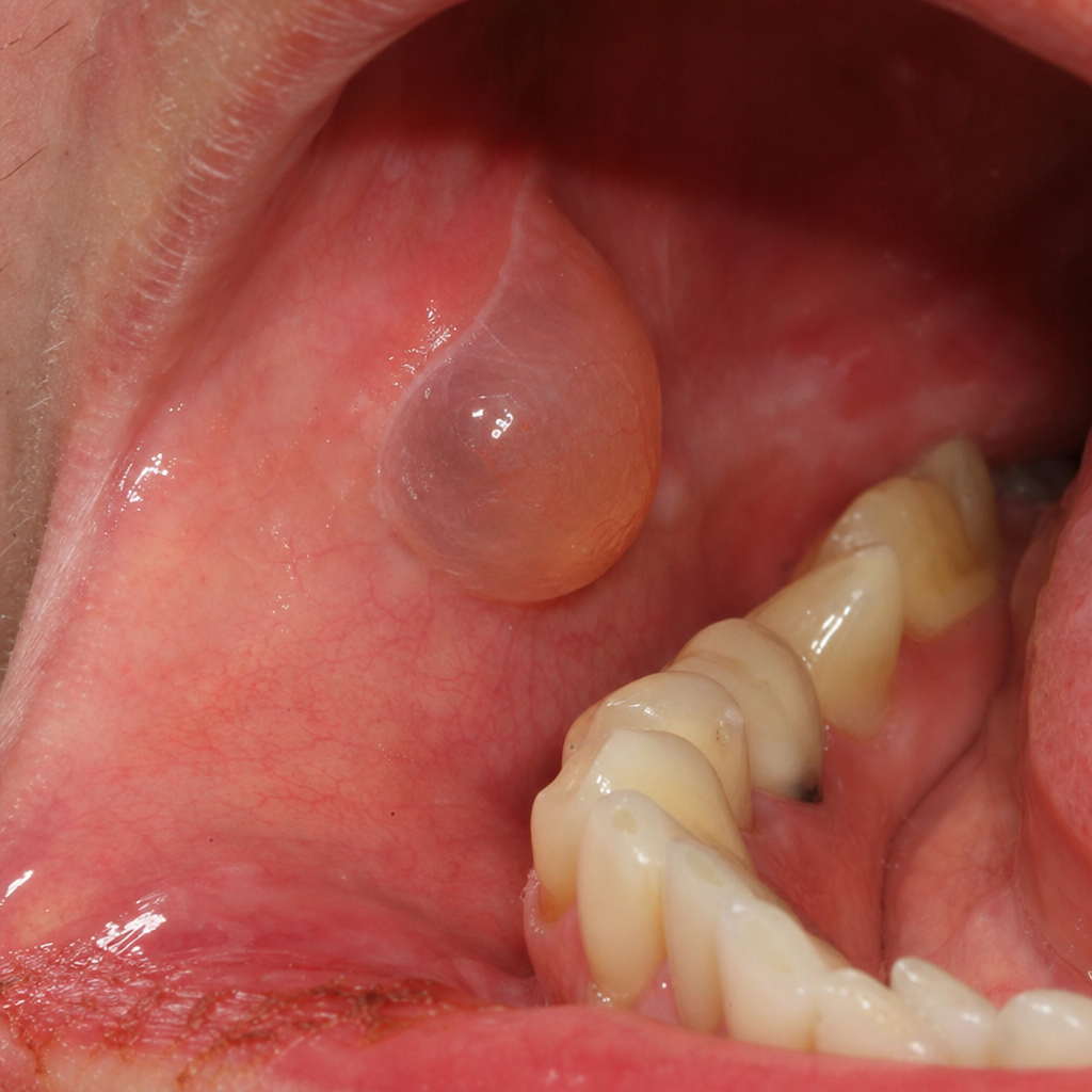
The following image shows a flaccid bulla. This finding is characteristically seen in:

Which of the following is NOT associated with erythema nodosum?
Acantholysis is seen in all except which of the following conditions?
Explanation: ***Subepidermal bulla*** - Pemphigus vulgaris is characterized by **intraepidermal bullae** resulting from acantholysis (loss of cohesion between keratinocytes), not subepidermal bullae. - **Subepidermal bullae** are characteristic of conditions like **bullous pemphigoid**, where the split occurs below the epidermis. *Positive Nikolsky’s sign* - The **Nikolsky's sign** is positive in pemphigus vulgaris, indicating the fragility of the skin where gentle lateral pressure causes epidermal shearing. - This sign is a direct result of the **intraepidermal blistering** due to weakened cell-to-cell adhesion. *Oral erosions* - **Oral erosions** are a very common and often the initial manifestation of pemphigus vulgaris, frequently preceding skin lesions. - These painful erosions are persistent and heal slowly, sometimes making eating difficult. *Tzanck smear showing acantholytic cells* - A **Tzanck smear** from a fresh blister in pemphigus vulgaris typically reveals **acantholytic cells**, which are detached, rounded keratinocytes with basophilic cytoplasm. - The presence of acantholytic cells confirms the **loss of intercellular adhesion** within the epidermis, a hallmark of pemphigus.
Explanation: ***Fishnet pattern of IgG*** - A **fishnet or reticular pattern** of **IgG deposition** on direct immunofluorescence (DIF) is characteristic of **pemphigus vulgaris**, indicating antibodies targeting **desmoglein 1 and 3** in the intracellular spaces of the epidermis. - This pattern corresponds to the **acantholysis** observed on biopsy, where loss of cell adhesion leads to intraepidermal blistering. *Linear IgA deposits* - **Linear IgA deposits** at the **dermal-epidermal junction** are characteristic of **linear IgA bullous dermatosis**, a blistering disorder distinct from pemphigus. - This pattern signifies **antibodies targeting components of the basement membrane zone**, not intraepidermal desmogleins. *Granular IgG deposits* - **Granular IgG deposits** in the skin are typically seen in conditions like **lupus erythematosus** or **dermatitis herpetiformis** when IgA is targeted, signifying immune complex deposition or specific antigen targeting. - This pattern is not associated with the pathogenesis of pemphigus vulgaris, which involves antibodies against desmosomal proteins. *Linear C3 deposits* - **Linear C3 deposits**, particularly at the **dermal-epidermal junction**, are a hallmark of **bullous pemphigoid**, often accompanied by linear IgG or IgA. - This indicates **complement activation** at the basement membrane zone, leading to subepidermal blistering, not the intraepidermal blistering seen in pemphigus vulgaris.
Explanation: ***Tzanck smear from the floor of bulla*** - A Tzanck smear from the floor of a bulla will reveal **acantholytic cells** (rounded keratinocytes that have lost their intercellular connections), which are characteristic of pemphigus, consistent with recurrent oral ulcers, denuded skin, and flaccid vesiculobullous eruptions. - This **bedside test** provides a rapid diagnosis by demonstrating the cytological features of acantholysis, differentiating it from other blistering disorders. *Gram staining of blister fluid* - This test is primarily used to identify **bacterial infections** and would show the morphology and Gram-staining characteristics of any bacteria present. - It would not provide information about the **acantholysis** or autoimmune nature of the blistering condition described. *Culture and sensitivity of blister fluid* - This investigation identifies **specific bacterial pathogens** and their antibiotic susceptibilities, which is useful for treating bacterial infections. - It would not help in diagnosing **autoimmune blistering diseases** like pemphigus, where bacteria are not the primary cause of the lesions. *Skin biopsy with immunofluorescence* - While a **skin biopsy with direct immunofluorescence** is the gold standard for confirming pemphigus by detecting autoantibodies, it is an **invasive procedure** requiring laboratory processing and is not considered a rapid bedside investigation. - The question specifically asks for the "most important **bed-side investigation**" helpful in establishing the diagnosis rapidly.
Explanation: ***Oral lesions may be found in any region, especially in the attached gingiva; ocular lesions can lead to blindness if untreated.*** - **Mucous membrane pemphigoid (MMP)** frequently manifests in the **oral cavity**, with the attached gingiva being the most common site, often presenting as **desquamative gingivitis**. - **Ocular involvement** occurs in 60-70% of cases and is a critical feature that can cause conjunctival scarring, symblepharon formation, ankyloblepharon, and eventually **blindness** if not recognized and managed early. - This statement captures the two most clinically significant features of MMP: the characteristic oral presentation and the sight-threatening ocular complications. *It presents as multiple, painful ulcers preceded by bullae which form below the epithelium at the basement membrane.* - While MMP does involve **subepithelial blister formation** at the basement membrane zone (confirmed by immunofluorescence showing linear IgG and C3 deposition), the clinical presentation is typically **chronic erosions and desquamation** rather than acute multiple painful ulcers. - The bullae in MMP are often **tense and intact initially** but rupture easily, leaving **slow-healing erosions** rather than the acute ulcerative picture this option suggests. - This description might be more characteristic of **pemphigus vulgaris** (which has flaccid, painful oral ulcers from intraepithelial bullae). *It primarily affects young adults and children, with peak incidence in the 2nd to 3rd decade of life.* - This is **incorrect**. MMP predominantly affects **middle-aged to elderly adults**, with peak incidence in the **6th to 7th decade of life** (ages 50-70 years). - The disease is rare in children and young adults, making this statement factually inaccurate.
Explanation: ***Bullous pemphigoid*** - Presents with **tense bullae** on an erythematous base, typically in elderly patients, unlike the **umbilicated papules** seen in this image. - Involves **subepidermal blistering** with **linear IgG deposition** at the basement membrane zone, not the viral inclusions of Molluscum contagiosum. *Pemphigus vegetans* - A rare variant of pemphigus vulgaris characterized by **vegetating plaques and pustules** in intertriginous areas, not discrete umbilicated lesions. - Shows **intraepidermal acantholysis** with **suprabasal clefting**, histologically distinct from the viral cytopathic changes in Molluscum contagiosum. *Pemphigus vulgaris* - Presents with **flaccid bullae** and painful **mucosal erosions** due to **autoantibodies against desmoglein 1 and 3**. - The **Nikolsky sign** is positive, and lesions are erosive rather than the solid, pearl-like papules characteristic of Molluscum contagiosum. *Pemphigus erythematosus* - Features **erythematous, scaly, crusted lesions** primarily on the **face and upper trunk** with a butterfly distribution. - Combines features of **lupus erythematosus** and pemphigus foliaceus, showing superficial blistering unlike the viral papules in this case.
Explanation: ***Herpes simplex*** - Herpes simplex virus (HSV) classically presents with **grouped vesicles on an erythematous base**, which perfectly matches this clinical presentation. - In **children**, HSV commonly affects the **buttocks** through autoinoculation or direct contact, especially in the diaper area. - The lesions are typically **painful and pruritic**, and may be preceded by tingling or burning sensation. - Diagnosis is confirmed by **Tzanck smear** (multinucleated giant cells), **PCR**, or **viral culture**. - Treatment includes **acyclovir** or other antivirals, especially for severe or recurrent cases. *Dermatitis herpetiformis* - While DH does present with intensely pruritic, grouped vesicles on an erythematous base, it is **extremely rare in children** and typically presents in **adults (3rd-4th decade)**. - Classic sites include **extensor surfaces** (elbows, knees), scalp, and buttocks, but the pediatric presentation makes this diagnosis unlikely. - It is strongly associated with **celiac disease** and responds to **gluten-free diet** and **dapsone**. *Bullous impetigo* - Bullous impetigo presents with **flaccid bullae** that rupture to form **honey-colored crusts**, not grouped vesicles. - It is a **bacterial infection** caused by *Staphylococcus aureus* producing exfoliative toxin. - Common in **young children**, particularly in warm, humid conditions. *Pemphigus* - Pemphigus is **extremely rare in children** and causes **fragile bullae** that easily rupture, leading to erosions. - Typically affects **mucous membranes first** (oral cavity), then skin. - It is an **autoimmune blistering disease** with antibodies against desmoglein, causing intraepidermal acantholysis.
Explanation: ***Fishnet pattern of IgG*** - Pemphigus vulgaris is characterized by **autoantibodies** (predominantly **IgG**) directed against **desmogleins 1 and 3**, components of desmosomes in the epidermis. - This binding leads to the **loss of cell adhesion** (acantholysis) and results in the characteristic **net-like or "fishnet" pattern** of IgG positivity on direct immunofluorescence (DIF), outlining keratinocyte cell surfaces. *Linear IgA deposits* - **Linear IgA bullous dermatosis** is characterized by the presence of continuous linear deposits of IgA along the **dermoepidermal junction** on direct immunofluorescence. - This condition is clinically and immunologically distinct from pemphigus vulgaris, which involves **intraepidermal deposition of IgG**. *Granular IgG deposits* - **Granular IgG deposits** at the dermoepidermal junction are typically seen in conditions like **lupus erythematosus**, particularly in the form of a **lupus band test**, rather than in blistering diseases like pemphigus. - Pemphigus involves **intercellular IgG deposition** within the epidermis, not granular deposits at the junction. *Linear C3 deposits* - **Linear C3 deposits** (often accompanied by IgG) along the **dermoepidermal junction** are characteristic of **bullous pemphigoid**, another autoimmune blistering disease. - Unlike pemphigus vulgaris, bullous pemphigoid involves autoantibodies targeting hemidesmosomes, leading to subepidermal blistering rather than intraepidermal acantholysis.
Explanation: ***Pemphigus vulgaris*** - The image shows a **flaccid bulla** with purulent fluid, characteristic of **pemphigus vulgaris**. This condition is marked by autoantibodies against desmogleins 1 and 3, which are crucial for keratinocyte adhesion, leading to **intraepidermal blistering** and the **Nikolsky sign**. - The flaccid nature of the bulla, often leading to easy rupture and erosions, is a hallmark of superficial blistering in pemphigus vulgaris, caused by the **loss of cell-to-cell adhesion** within the epidermis. *Pemphigus vegetans* - This is a rare variant of pemphigus vulgaris characterized by **vegetating plaques** and **hyperkeratotic lesions**, particularly in intertriginous areas. - While it starts with bullae, the predominant feature is the development of fungating, vegetative lesions rather than the flaccid bulla seen here. *Pemphigus erythematosus* - Pemphigus erythematosus, also known as Senear-Usher syndrome, is considered a localized form of pemphigus foliaceus with features of **lupus erythematosus**. - It presents with **scaling, crusting, and erythematous lesions** resembling lupus, along with superficial bullae, typically on the face and scalp. *Bullous pemphigoid* - Bullous pemphigoid typically presents with **tense bullae** that are less prone to rupture, unlike the flaccid bulla shown in the image. - It is caused by autoantibodies against hemidesmosomal proteins (BP180 and BP230), resulting in **subepidermal blistering**, meaning the blister forms below the epidermis and is therefore more resilient.
Explanation: ***Pemphigus vulgaris*** - **Pemphigus vulgaris** is an **autoimmune blistering disease** that affects the skin and mucous membranes, characterized by flaccid bullae, not subcutaneous nodules. - Its pathophysiology involves **autoantibodies** against **desmoglein 1 and 3**, leading to **acantholysis**, which is distinct from the inflammatory changes seen in erythema nodosum. *Tuberculosis* - **Tuberculosis (TB)** is a common infectious cause of **erythema nodosum**, especially in regions with high TB prevalence. - The development of erythema nodosum in TB is often considered a **hypersensitivity reaction** to mycobacterial antigens. *Sarcoidosis* - **Sarcoidosis** is a systemic granulomatous disease, and **erythema nodosum** can be a prominent cutaneous manifestation, particularly in **Löfgren's syndrome**. - Its presence with **bilateral hilar lymphadenopathy** and **arthralgia** is highly suggestive of acute sarcoidosis. *Leprosy* - **Leprosy**, caused by *Mycobacterium leprae*, can be associated with **erythema nodosum leprosum (ENL)**, which is a type 2 lepra reaction. - **ENL** involves the formation of painful, tender, inflamed nodules that resemble erythema nodosum and is linked to elevated immune complex deposition.
Explanation: ***Bullous pemphigoid*** - This condition involves **subepidermal blistering**, meaning the separation of the epidermis from the dermis, which occurs *below* the **basal cell layer**. - **Acantholysis**, the loss of cohesion between keratinocytes *within* the epidermis, does not occur in bullous pemphigoid, making it the correct answer. *Pemphigus vulgaris* - This is an **autoimmune blistering disease** characterized by the presence of autoantibodies against **desmoglein 3** (and often desmoglein 1). - This leads to intraepidermal blistering caused by **acantholysis**, the primary pathophysiological event. *Darier's disease* - This is an **autosomal dominant genodermatosis** characterized by abnormal keratinization and acantholysis. - Due to defects in **ATP2A2** (encoding SERCA2), there is impaired calcium handling in keratinocytes, leading to premature desmosomal degradation and **acantholysis**. *SSSS (Staphylococcal Scalded Skin Syndrome)* - Caused by **exfoliative toxins** (ETA and ETB) produced by *Staphylococcus aureus* that target **desmoglein 1**. - The cleavage of desmoglein 1 results in superficial **intraepidermal blistering** due to **acantholysis** in the granular layer of the epidermis.
Get full access to all questions, explanations, and performance tracking.
Start For Free