Best site for taking biopsy for HSV esophagitis is:
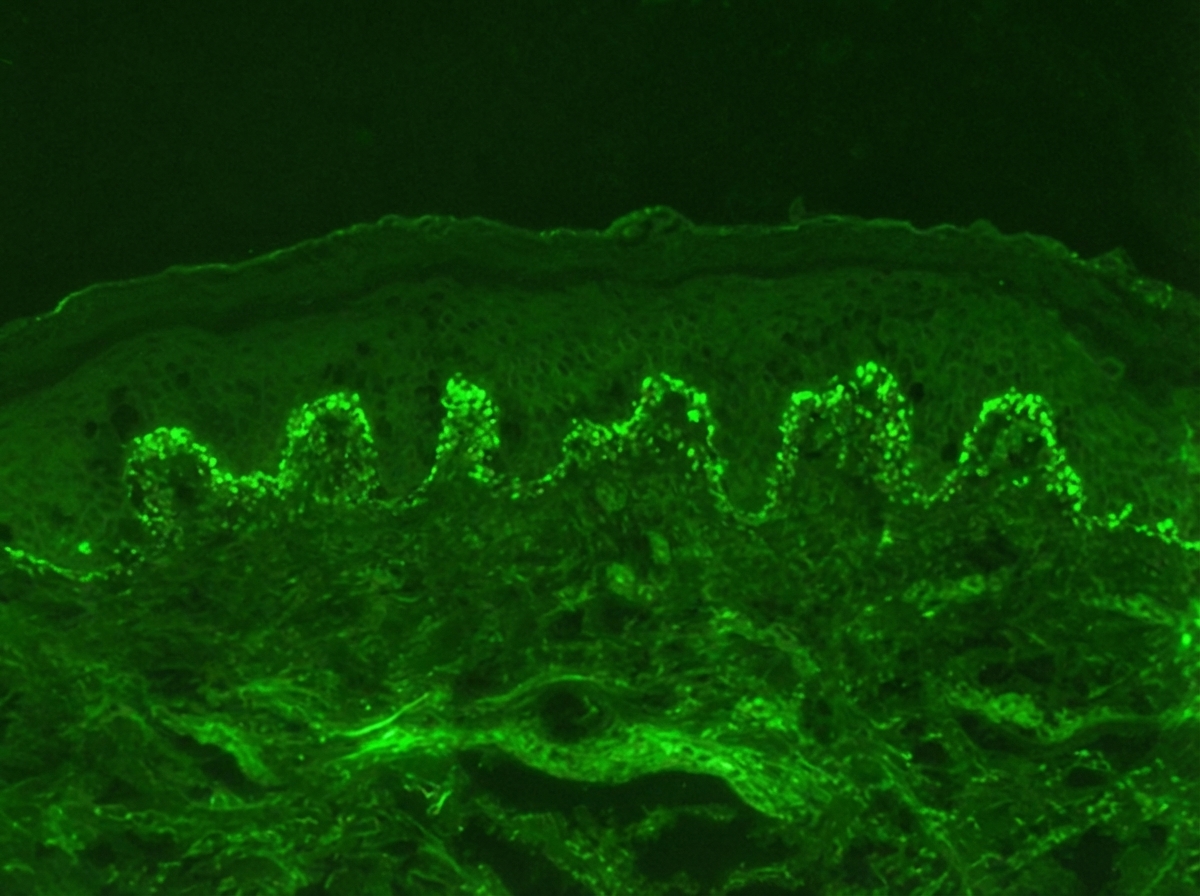
Identify the diagnosis based on the dermatology immunofluorescence (IF) image provided.

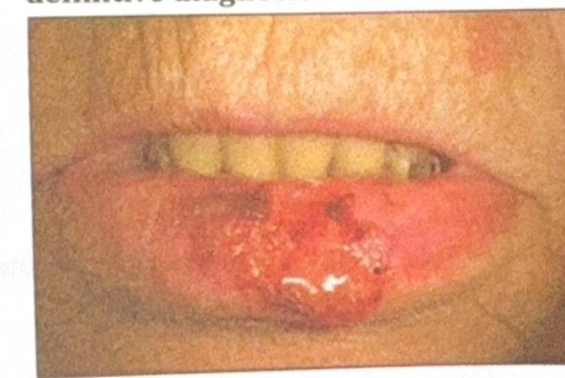
An elderly patient presents with a non-healing ulcerative lesion on the lower lip, as shown in the image. The lesion has been gradually enlarging over the past few months. Suspecting squamous cell carcinoma (SCC), what is the most appropriate method to obtain a biopsy for definitive diagnosis?

A patient presents with cutaneous vasculitis, glomerulonephritis and peripheral neuropathy. What investigation is to be performed next for work up?
Acute febrile neutrophilic dermatosis is seen in-
Which of the following statements about actinic lichen planus is false?
Which of the following is a xenodiagnostic method?
A skin biopsy shows suprabasilar acantholysis with 'row of tombstones' appearance. Which immunofluorescence pattern would confirm pemphigus vulgaris?
A child was diagnosed as a case of pauci-immune crescentic glomerulonephritis. The treatment to be given in this child is –
Patient presenting with cutaneous vasculitis, glomerulonephritis, peripheral neuropathy, which investigation is to be performed next that will help you diagnose the condition?
Explanation: ***Edge of ulcer*** - The **edge of the ulcer** is the preferred site for biopsy in HSV esophagitis because it is where the **actively replicating viral particles** and **cytopathic effects** are most likely to be found. - This area allows for the detection of characteristic **ground-glass nuclei**, **Cowdry type A inclusions**, and **multinucleated giant cells** indicative of HSV infection. *Base of ulcer* - While the base of the ulcer might show inflammatory changes, it is less likely to contain actively replicating virus or viable host cells exhibiting the classic **cytopathic changes** seen in HSV. - The base often consists of **necrotic debris** and granulation tissue, making a definitive diagnosis more difficult. *Adjacent indurated area around ulcer* - An indurated area around an ulcer could suggest chronic inflammation or other pathologies, but for acute HSV infection, it is less likely to yield diagnostic viral effects. - This region may show secondary inflammatory changes rather than the primary viral effects at the site of invasion. *Surrounding normal mucosa* - The normal mucosa surrounding the ulcer is unlikely to show any direct histological evidence of HSV infection. - Biopsying this area would not be diagnostic as the virus primarily affects and ulcerates the epithelial lining.
Explanation: ***Dermatitis herpetiformis*** - The immunofluorescence image shows **granular IgA deposits** at the **dermal papillae region**, which is characteristic of dermatitis herpetiformis. - This condition is strongly associated with **celiac disease** and presents with intensely pruritic papules and vesicles. *Pemphigus vulgaris* - Immunofluorescence in pemphigus vulgaris typically shows a **fishnet pattern** of IgG deposits throughout the **epidermis**, reflecting antibodies against desmoglein 3 and 1. - This pattern is an intercellular deposition, not granular at the dermal papillae. *Pemphigus foliaceus* - Similar to pemphigus vulgaris, pemphigus foliaceus also exhibits **intercellular IgG deposits** in the epidermis, but it is usually more superficial, targeting desmoglein 1. - The image does not show this intercellular epidermal staining. *Bullous pemphigoid* - Bullous pemphigoid is characterized by **linear IgG and C3 deposits along the dermal-epidermal junction** (basement membrane zone). - The image distinctly shows granular IgA, not linear IgG/C3, and specifically in the dermal papillae.
Explanation: ***Incisional*** - An **incisional biopsy** is the most appropriate method for obtaining a definitive diagnosis of suspected squamous cell carcinoma (SCC) of the lip. - This technique involves removing a **wedge-shaped portion of the lesion** that includes both the tumor tissue and a margin extending into normal tissue, with adequate depth to assess invasion. - Incisional biopsy provides sufficient tissue for **histopathological examination**, including assessment of tumor grade, depth of invasion, and other prognostic factors critical for staging and treatment planning. - For larger or suspicious lesions where complete excision might cause significant cosmetic deformity, incisional biopsy allows for **diagnosis confirmation before definitive surgical management**. *Superficial biopsy from the border with normal tissue* - A superficial or shave biopsy is **inadequate for SCC diagnosis** as it does not provide information about the depth of invasion, which is crucial for staging and prognosis. - Squamous cell carcinoma requires assessment of invasion into underlying dermis and deeper structures, which cannot be evaluated with superficial sampling. - Superficial biopsies may lead to **underdiagnosis** or incomplete staging, potentially compromising treatment planning. *Excisional* - While excisional biopsy (complete removal with margins) can be appropriate for **small, well-defined lesions** (<1 cm), it may not be the first choice for larger or gradually enlarging lesions. - Complete excision without prior histological confirmation might result in **inadequate margins** if malignancy is confirmed, requiring re-excision. - For lip lesions, unnecessary wide excision can cause **significant cosmetic and functional defects** if the lesion proves benign or requires specialized reconstruction. *Deep tissue biopsy* - This is not standard terminology in the context of lip lesions and lacks specificity regarding the sampling technique. - The term "deep tissue biopsy" is more commonly used for suspected soft tissue tumors or deep-seated lesions, not for mucocutaneous SCC.
Explanation: **ANCA** - The combination of **cutaneous vasculitis**, **glomerulonephritis**, and **peripheral neuropathy** points strongly towards a Systemic Vasculitis [1]. **ANCA** (anti-neutrophil cytoplasmic antibodies) are crucial for diagnosing and classifying **ANCA-associated vasculitides**, such as **Granulomatosis with Polyangiitis (GPA)**, **Microscopic Polyangiitis (MPA)**, and **Eosinophilic Granulomatosis with Polyangiitis (EGPA)**, which perfectly fit this clinical picture [1]. - Identification of specific ANCA patterns (p-ANCA or c-ANCA) and their corresponding target antigens (e.g., **MPO** or **PR3**) is essential for confirming the diagnosis and guiding treatment. *HBsAg* - While **Hepatitis B virus (HBV) infection** can cause **polyarteritis nodosa (PAN)**, which may present with vasculitis and neuropathy, it typically does not involve **glomerulonephritis** as a primary feature [1]. - Testing for HBsAg would be relevant if PAN was strongly suspected or if other risk factors for HBV were present, but ANCA testing is more direct for the given clinical triad. *RA factor* - **Rheumatoid factor (RF)** is primarily associated with **Rheumatoid Arthritis**, which is an inflammatory joint disease and does not typically present with this triad of **vasculitis**, **glomerulonephritis**, and **neuropathy** [2]. - Although some connective tissue diseases can have vasculitic manifestations, RF itself is not a primary diagnostic marker for systemic vasculitis. *CRP* - **C-reactive protein (CRP)** is a general **marker of inflammation**, which would undoubtedly be elevated in a patient with systemic vasculitis [1]. - However, CRP is **non-specific** and would not help in narrowing down the diagnosis or determining the etiology of the vasculitis, making it less useful for specific workup compared to ANCA.
Explanation: ***Sweet syndrome*** - Sweet syndrome is also known as **Acute febrile neutrophilic dermatosis**. - It presents with **fever**, painful erythematous plaques, and a prominent **neutrophilic infiltrate** in the dermis. *Behcet's syndrome* - Behcet's syndrome is characterized by **recurrent oral and genital ulcers**, uveitis, and skin lesions like **erythema nodosum** or **pathergy**. - It is a systemic vasculitis, not typically presenting as acute febrile neutrophilic dermatosis. *Haberman syndrome* - Haberman syndrome is better known as **Pityriasis lichenoides et varioliformis acuta (PLEVA)**. - It is a rare skin condition characterized by an acute eruption of **papules** and **erosions** that can resemble **chickenpox**. *Kasabach-Merritt syndrome* - This syndrome is a rare disorder involving **vascular tumors** (e.g., hemangiomas) that lead to profound **thrombocytopenia** and **consumptive coagulopathy**. - It does not present with acute febrile neutrophilic dermatosis.
Explanation: ***Associated with severe pruritus*** - While other forms of lichen planus, especially the typical cutaneous type, are often associated with **severe pruritus**, actinic lichen planus is typically characterized by **mild or absent pruritus**. - Its presentation is often more focused on pigmentary changes and papules in sun-exposed areas rather than intense itching. *Autoimmune etiology* - **Lichen planus**, including its actinic variant, is recognized as an **autoimmune disease**. - It involves a **T-cell-mediated immune response** against basal keratinocytes. *Violaceous brown papules* - Actinic lichen planus commonly presents with **violaceous, brown, or hyperpigmented papules and plaques**. - These lesions often exhibit a subtle **annular or reticulated pattern**. *Usually affects exposed areas of body* - As its name suggests ("actinic" referring to light), this variant of lichen planus preferentially affects **sun-exposed areas** such as the face, neck, and dorsal hands. - This distribution distinguishes it from classic lichen planus, which can occur anywhere but often affects the flexor surfaces of the wrists, ankles, and oral mucosa.
Explanation: ***Injecting Aedes thorax with blood of a suspected dengue patient*** - **Xenodiagnosis** involves using a live arthropod vector (such as an *Aedes* mosquito) to detect the presence of pathogens in a host by feeding the vector on the host and subsequently examining the vector for infection. - In this method, the mosquito acts as a biological incubator or amplifier for the dengue virus, which can then be detected within the mosquito, indicating infection in the patient. *Intradermal test on guinea pigs for toxigenicity of Corynebacterium diphtheria* - This is an **animal pathogenicity test** to determine if the *Corynebacterium diphtheriae* strain produces toxin, but it is not xenodiagnosis as the animal is the test subject, not a vector for detection. - The test assesses the virulence of the bacterium directly in the animal, rather than using the animal to detect an existing infection in another host. *Injecting a hamster with splenic biopsy for diagnosis of leishmaniasis* - This method is an example of **animal inoculation** or **culture in vivo**, where a susceptible animal is used to grow and amplify a suspected pathogen from a patient sample. - While it uses an animal for diagnosis, it's not xenodiagnosis because the hamster is not a natural vector that feeds on the patient to acquire the pathogen. *Rabbit ileal loop for enterotoxigenic Escherichia coli* - This is an **animal model** used to detect the production of **enterotoxins** by *Escherichia coli*, which cause fluid accumulation in the rabbit ileum. - This method tests for the pathogenic effect of the bacteria rather than using an arthropod vector to detect infection from a human host.
Explanation: ***Fishnet pattern of IgG*** - Pemphigus vulgaris is characterized by **autoantibodies** (predominantly **IgG**) directed against **desmogleins 1 and 3**, components of desmosomes in the epidermis. - This binding leads to the **loss of cell adhesion** (acantholysis) and results in the characteristic **net-like or "fishnet" pattern** of IgG positivity on direct immunofluorescence (DIF), outlining keratinocyte cell surfaces. *Linear IgA deposits* - **Linear IgA bullous dermatosis** is characterized by the presence of continuous linear deposits of IgA along the **dermoepidermal junction** on direct immunofluorescence. - This condition is clinically and immunologically distinct from pemphigus vulgaris, which involves **intraepidermal deposition of IgG**. *Granular IgG deposits* - **Granular IgG deposits** at the dermoepidermal junction are typically seen in conditions like **lupus erythematosus**, particularly in the form of a **lupus band test**, rather than in blistering diseases like pemphigus. - Pemphigus involves **intercellular IgG deposition** within the epidermis, not granular deposits at the junction. *Linear C3 deposits* - **Linear C3 deposits** (often accompanied by IgG) along the **dermoepidermal junction** are characteristic of **bullous pemphigoid**, another autoimmune blistering disease. - Unlike pemphigus vulgaris, bullous pemphigoid involves autoantibodies targeting hemidesmosomes, leading to subepidermal blistering rather than intraepidermal acantholysis.
Explanation: ***Prednisolone + Cyclophosphamide*** - **Pauci-immune crescentic glomerulonephritis** (also known as ANCA-associated vasculitis) is a rapidly progressive and severe autoimmune condition affecting the kidneys. - **Combination therapy** with a corticosteroid (like prednisolone) and an immunosuppressant (like cyclophosphamide) is the standard and most effective initial treatment to control inflammation and suppress the immune response. *Immunoglobulins* - **Intravenous immunoglobulins (IVIG)** are sometimes used in autoimmune diseases, but they are not the first-line treatment for pauci-immune crescentic glomerulonephritis due to its aggressive nature. - IVIG might be considered as an **adjunctive therapy** or in specific cases of refractory disease, but not as monotherapy. *Cyclophosphamide* - While **cyclophosphamide** is a critical component of treatment for **pauci-immune crescentic glomerulonephritis** due to its potent immunosuppressive effects, it is not used alone. - It is typically combined with a **corticosteroid** (such as prednisolone) to achieve a more rapid and comprehensive anti-inflammatory and immunosuppressive effect. *Methylprednisolone* - **Methylprednisolone** (a corticosteroid) is routinely used in the initial management of **pauci-immune crescentic glomerulonephritis** to rapidly reduce inflammation. - However, it is generally part of a **combination regimen** and is not sufficient as monotherapy for inducing remission in this severe form of glomerulonephritis.
Explanation: ***ANCA*** - The combination of **cutaneous vasculitis**, **glomerulonephritis**, and **peripheral neuropathy** strongly suggests a **systemic vasculitis**, for which ANCA testing is crucial for diagnosis and classification [1]. - **ANCA (anti-neutrophil cytoplasmic antibodies)** are primarily associated with **ANCA-associated vasculitides** such as **granulomatosis with polyangiitis (GPA)**, **microscopic polyangiitis (MPA)**, and **eosinophilic granulomatosis with polyangiitis (EGPA)** [1]. *HbsA* - **Hepatitis B surface antigen (HbsAg)** testing is relevant in the context of **polyarteritis nodosa (PAN)**, a type of vasculitis sometimes associated with hepatitis B infection. - However, the clinical presentation here, particularly the glomerulonephritis, is less typical for PAN and more suggestive of ANCA-associated vasculitis. *RA factor* - **Rheumatoid factor (RF)** is primarily associated with **rheumatoid arthritis**, which typically presents with inflammatory polyarthritis rather than widespread vasculitis, glomerulonephritis, and peripheral neuropathy. - While some cases of **rheumatoid vasculitis** can occur, the constellation of symptoms points more strongly towards a primary systemic vasculitis. *Complement levels (C3, C4)* - **Low complement levels (C3, C4)** are characteristic of **cryoglobulinemic vasculitis** (often associated with **HCV infection**), **lupus nephritis**, or certain forms of **post-streptococcal glomerulonephritis**. - While vasculitis and glomerulonephritis can occur in these conditions, the specific combination presented (cutaneous vasculitis, glomerulonephritis, peripheral neuropathy) makes **ANCA-associated vasculitis** a more direct and urgent consideration for initial investigation.
Get full access to all questions, explanations, and performance tracking.
Start For Free