Which of the following statements regarding collagen synthesis is incorrect?
Ubiquitin is involved in what process?
Carboxylation of clotting factors by vitamin K is required for them to be biologically active. Which amino acid is carboxylated?
What is the major site of protein glycosylation?
Which of the following is not a characteristic feature of Alzheimer's disease?
Which of the following is considered a poor prognostic marker in multiple myeloma (MM)?
Statement 1 - A 59-year-old patient presents with flaccid bullae. Histopathology shows a suprabasal acantholytic split. Statement 2 - The row of tombstones appearance is diagnostic of Pemphigus vulgaris.
During eukaryotic protein synthesis, phosphorylation of which of the following is enhanced by insulin?
Protein segregation occurs in which organelle?
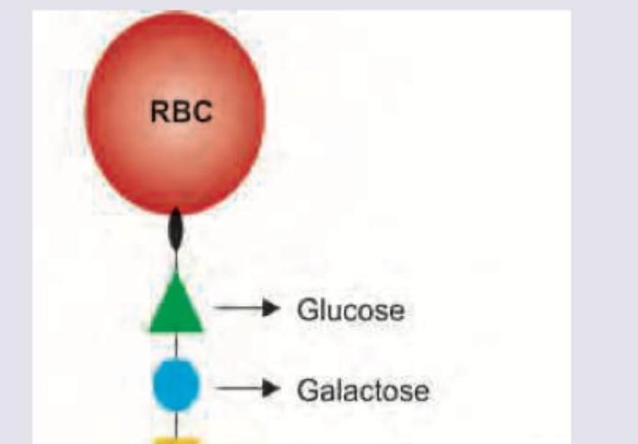
Name the antigen marked as X determining blood group A.

Explanation: ***Hydroxylation of proline occurs in Golgi apparatus*** - This statement is incorrect because the **hydroxylation of proline** residues occurs in the **endoplasmic reticulum** (ER), not the Golgi apparatus. - This step is critical for forming stable **triple helix** structures of collagen and requires **vitamin C**. *Synthesized in ribosomes as preprocollagen* - This statement is correct. Collagen synthesis begins in the cytoplasm, where mRNA is translated by **ribosomes** into **preprocollagen**, which contains a signal peptide. - The signal peptide directs the nascent polypeptide chain into the lumen of the **endoplasmic reticulum**. *Hydroxylation of lysine occurs in ER* - This statement is correct. Following entry into the ER, specific **lysine** residues are hydroxylated by **lysyl hydroxylase** to form hydroxylysine. - This hydroxylation, along with that of proline, is crucial for **cross-linking** and stability of the collagen molecule. *Triple helix assembly occurs in ER* - This statement is correct. After hydroxylation and glycosylation of some residues, three procollagen alpha chains self-assemble to form a **triple helix** within the **endoplasmic reticulum**. - This assembly is stabilized by **disulfide bonds** at the C-terminal ends and molecular chaperones.
Explanation: ***Protein degradation*** - **Ubiquitin** is a small regulatory protein that attaches to other proteins as a signal, primarily for their **degradation** by the **proteasome**. - This process, known as **ubiquitination**, marks misfolded, damaged, or no longer needed proteins for targeted destruction. *Protein folding* - This process is primarily mediated by **chaperone proteins**, which assist in the correct three-dimensional structuring of polypeptides. - While ubiquitin can sometimes influence protein folding indirectly by marking misfolded proteins for degradation, its direct role is not in the folding itself. *Synthesis of nucleic acid* - The synthesis of **nucleic acids** (DNA and RNA) is carried out by **DNA polymerases** and **RNA polymerases**, respectively. - Ubiquitin is not involved in the enzymatic processes of replication or transcription. *Glycosylation of proteins* - **Glycosylation** is the enzymatic addition of carbohydrate moieties to proteins, typically occurring in the **endoplasmic reticulum** and **Golgi apparatus**. - This process is crucial for protein function, trafficking, and cell-cell recognition, but ubiquitin has no direct role in it.
Explanation: ***Glutamate*** - **Vitamin K** acts as a cofactor for the enzyme **gamma-glutamyl carboxylase**, which carboxylates specific **glutamate residues** in clotting factors (II, VII, IX, and X). - This carboxylation forms **gamma-carboxyglutamate (Gla)** residues, which allows the clotting factors to bind **calcium ions** and thereby to phospholipid surfaces, enabling the coagulation cascade. *Histidine* - **Histidine** is an amino acid that plays a role in enzyme active sites and metal ion chelation, but it is not the target for **vitamin K-dependent carboxylation**. - Its imidazole ring can donate and accept protons, giving it a role in **pH buffering** and catalytic mechanisms. *Serine* - **Serine** is an amino acid that undergoes post-translational modifications such as **phosphorylation** (by kinases) and **O-glycosylation**. - However, it is not the amino acid specifically targeted by **vitamin K-dependent carboxylation** for the activation of clotting factors. *Aspartate* - **Aspartate** is an acidic amino acid that can bind **metal ions** and participates in various metabolic pathways and enzyme active sites. - While structurally similar to glutamate, it is not the amino acid specifically targeted by **vitamin K-dependent carboxylation** for the activation of clotting factors.
Explanation: ***ER and Golgi body*** - The **endoplasmic reticulum (ER)** is the primary site for **N-linked glycosylation**, where carbohydrates are added to the asparagine residues of nascent proteins. - The **Golgi apparatus** is crucial for further modification and processing of these N-linked glycans, as well as the site for **O-linked glycosylation**, where sugars are added to serine or threonine residues. *Ribosome and Golgi body* - **Ribosomes** are responsible for **protein synthesis (translation)** but do not directly perform glycosylation, which is a post-translational modification. - While the **Golgi body** is a site of glycosylation, the ribosome's inclusion makes this option incorrect as the ribosome's role precedes glycosylation. *ER and Ribosome* - The **ER** is a major site of protein glycosylation, especially N-linked glycosylation. - However, **ribosomes** are involved in protein synthesis and lack the enzymatic machinery for adding sugar moieties to proteins. *Ribosome and Cytoplasm* - **Ribosomes** synthesize proteins, but glycosylation does not occur there. - The **cytoplasm** is the site for many metabolic pathways, but major protein glycosylation events mostly occur within the ER and Golgi.
Explanation: ***Correct Answer: Lewy bodies*** - **Lewy bodies** are abnormal aggregates of protein, primarily **alpha-synuclein**, found in the brains of individuals with **Parkinson's disease** and **Lewy body dementia**, but **not typically in Alzheimer's disease** [1]. - While there can be overlap in pathologies, the presence of Lewy bodies as a primary feature suggests a different neurodegenerative process than Alzheimer's. - This is the correct answer because it is **NOT a characteristic feature** of Alzheimer's disease. *Incorrect: Neurofibrillary tangles* - **Neurofibrillary tangles** are intracellular aggregates of hyperphosphorylated **tau protein** and are a **hallmark pathological feature** of Alzheimer's disease. - These tangles disrupt neuronal function and axonal transport, leading to neuronal death. - This IS a characteristic feature of Alzheimer's disease. *Incorrect: Senile (neuritic) plaques* - **Senile plaques**, also known as **neuritic plaques**, are extracellular deposits of **beta-amyloid protein** that are a **characteristic pathological feature** of Alzheimer's disease. - These plaques accumulate outside neurons, leading to inflammation and neuronal dysfunction. - This IS a characteristic feature of Alzheimer's disease. *Incorrect: Amyloid Angiopathy* - **Cerebral amyloid angiopathy** (CAA) is the deposition of **amyloid-beta protein** in the walls of small and medium-sized blood vessels in the central nervous system, particularly the cerebral cortex and leptomeninges. - It is present in **over 90% of Alzheimer's disease cases** and is a significant contributor to cognitive decline and hemorrhagic events in these patients. - This IS a characteristic feature of Alzheimer's disease. **References:** [1] Kumar V, Abbas AK, et al.. Robbins and Cotran Pathologic Basis of Disease. 9th ed. The Central Nervous System, pp. 1296-1298.
Explanation: ***B2 microglobulins*** - Elevated levels of **B2 microglobulin** are a significant indicator of increased tumor burden and are used in the **International Staging System (ISS)** for multiple myeloma, correlating with shorter survival [1]. - This protein is present on the surface of most nucleated cells and its accumulation reflects **renal impairment** or increased cell turnover. *Serum Creatinine* - While elevated **serum creatinine** can indicate **renal insufficiency**, a common complication in MM, it is not a direct measure of tumor burden or aggression in the same way as B2 microglobulin [1]. - **Renal failure** can be a poor prognostic factor in MM, but creatinine itself is a marker of organ damage rather than disease progression or malignancy. *Hypercalcemia* - **Hypercalcemia** is a common complication in MM due to increased **bone resorption**, but it is generally manageable and not considered a primary prognostic marker for disease aggressiveness itself. - While severe hypercalcemia can contribute to overall morbidity, its presence doesn't directly stage the disease or predict survival as significantly as B2 microglobulin. *Telomerase* - **Telomerase** is an enzyme involved in maintaining telomere length and is often overexpressed in various cancers, including MM, allowing cells to proliferate indefinitely. - While it plays a role in the **pathogenesis** of MM, its utility as a prognostic marker in routine clinical practice is not as established or as widely used as B2 microglobulin.
Explanation: ***Correct: Statements 1 & 2 are correct, 2 is not explaining 1*** **Analysis of Statement 1:** - A 59-year-old patient with **flaccid bullae** and **suprabasal acantholytic split** on histopathology is the classic presentation of **Pemphigus vulgaris** - The flaccid (easily ruptured) nature of bullae distinguishes it from tense bullae seen in bullous pemphigoid - The suprabasal location of the split (just above the basal layer) with acantholysis (loss of cell-to-cell adhesion) is pathognomonic - **Statement 1 is CORRECT** ✓ **Analysis of Statement 2:** - The **"row of tombstones" or "tombstone appearance"** is indeed a diagnostic histopathological feature of Pemphigus vulgaris - This appearance results from basal keratinocytes remaining attached to the basement membrane while suprabasal cells separate due to acantholysis - The intact basal cells standing upright resemble a row of tombstones - **Statement 2 is CORRECT** ✓ **Does Statement 2 explain Statement 1?** - Statement 2 describes a **histopathological appearance** (tombstone pattern) that is a **consequence** of the suprabasal split - However, it does NOT explain the **underlying cause** of the flaccid bullae or the suprabasal split - The true explanation involves **IgG autoantibodies against desmoglein 3 (and desmoglein 1)**, which attack intercellular adhesion structures (desmosomes), causing **acantholysis** - Therefore, **Statement 2 does NOT explain Statement 1** ✗ *Incorrect: Statement 2 is the correct explanation for Statement 1* - While both statements describe features of Pemphigus vulgaris, the tombstone appearance is a descriptive finding, not an explanatory mechanism *Incorrect: Statements 1 and 2 are incorrect* - Both statements are medically accurate descriptions of Pemphigus vulgaris features *Incorrect: Statement 1 is incorrect* - Statement 1 correctly describes the cardinal clinical and histopathological features of Pemphigus vulgaris
Explanation: ***eIF4E*** - Insulin activates the **mTOR pathway**, which leads to activation of **Mnk1/2 kinases** that phosphorylate eIF4E at **Ser209**. - This phosphorylation enhances eIF4E's **affinity for the 5' cap structure** and increases **cap-dependent translation initiation** efficiency. *eIF4G* - While eIF4G is essential for **eIF4F complex formation**, its phosphorylation is not the primary target enhanced by insulin signaling. - Insulin's effect on eIF4G is mainly **indirect through 4E-BP1 phosphorylation**, which releases eIF4E to bind eIF4G. *eIF2* - **eIF2 phosphorylation** by kinases like **PERK, PKR, and GCN2** inhibits translation initiation during stress conditions. - This is **opposite to insulin's anabolic effects**, as insulin signaling typically promotes conditions that reduce eIF2 phosphorylation. *eIF4A* - eIF4A functions as an **RNA helicase** in the eIF4F complex, unwinding mRNA secondary structures. - While important for translation, **direct phosphorylation enhancement by insulin** is not a primary mechanism for eIF4A regulation.
Explanation: ***Golgi apparatus*** - The **Golgi apparatus** is a central organelle for **protein modification, sorting, and packaging** into vesicles for delivery to various cellular destinations. - It acts as a "post office" of the cell, directing proteins to their correct locations through **segregation** into specific secretory or transport pathways. *Peroxisomes* - **Peroxisomes** are involved in **metabolic processes** such as fatty acid oxidation and detoxification. - While they import some proteins, their primary role is not in the overall **segregation** and trafficking of proteins for diverse cellular destinations. *ER* - The **endoplasmic reticulum (ER)** is where proteins are synthesized (rough ER) and undergo initial folding and modification, including glycosylation. - However, the ER's main function is protein synthesis and early modification, not the final **segregation** and sorting for transport to different cellular locations. *Mitochondria* - **Mitochondria** are primarily responsible for **ATP production** through cellular respiration and houses its own genome. - While mitochondria import specific proteins necessary for their function, they are not involved in the general **segregation** of proteins destined for other organelles or secretion.
Explanation: ***N-Acetyl-Galactosamine*** - Blood group A antigens are formed by the addition of **N-acetylgalactosamine** to the H antigen precursor molecule on the surface of red blood cells. - This sugar modification is catalyzed by the **A transferase enzyme**, which is specific for N-acetylgalactosamine. *N-Acetyl-Glucosamine* - While N-acetylglucosamine is a component of many glycans, it is not the terminal sugar that defines the **blood group A antigen**. - **N-acetylglucosamine** is a key building block for the H antigen and other blood group precursors, but not the specific modifying sugar for A. *Dermatan sulphate* - **Dermatan sulfate** is a **glycosaminoglycan** primarily found in connective tissues, skin, and blood vessels. - It plays a role in wound healing and coagulation, but is not involved in **ABO blood group determination**. *Keratan sulfate* - **Keratan sulfate** is another **glycosaminoglycan** found in cartilage, cornea, and bone. - It contributes to tissue hydration and structural integrity, but it is not part of the **ABO blood group antigens**.
Get full access to all questions, explanations, and performance tracking.
Start For Free