
Protein Structure and Function — MCQs

On this page
What is the primary protein that binds to thyroxine?
A patient has a long arm span, hypermobile joints and ectopia lentis. What is the defective protein?
Which of the following amino acids primarily acts as a buffer in blood due to its ability to accept and donate protons at physiological pH?
Which among the following helps in the maturation of collagen?
IGF-1 and IGF-2 are structurally most similar to which of the following molecules?
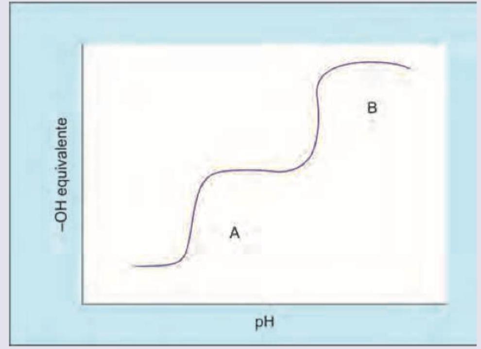
What is the pH of the zwitterion as shown in the titration curve of glycine?
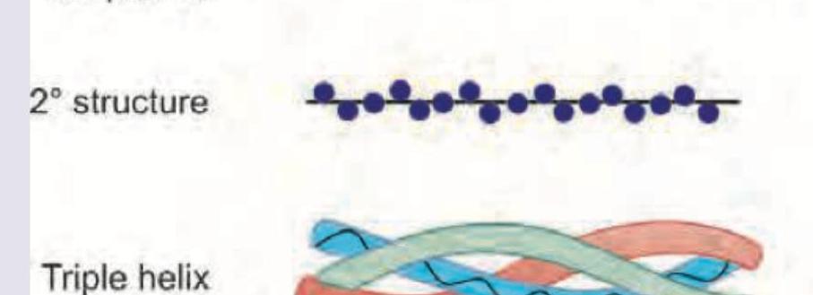
All are correct about the protein shown in the figure except? (Recent NEET Pattern 2016-17)
All of the statements are true about the titration curve of a protein except:
Match the following molecules with their carriers in plasma: MOLECULES: 1. Thyroxine 2. Fatty acid 3. Hemoglobin 4. Heme CARRIERS: A. Hemopexin B. Prealbumin C. Albumin D. Haptoglobin
Beta hCG is structurally similar to which biochemical moiety?
Practice by Chapter
Amino Acids: Structure and Properties
Practice Questions
Peptide Bond Formation
Practice Questions
Primary Structure of Proteins
Practice Questions
Secondary Structure of Proteins
Practice Questions
Tertiary and Quaternary Structures
Practice Questions
Protein Folding and Chaperones
Practice Questions
Protein Domains and Motifs
Practice Questions
Structure-Function Relationships
Practice Questions
Hemoglobin and Myoglobin
Practice Questions
Collagen and Elastin
Practice Questions
Albumin and Plasma Proteins
Practice Questions
Post-Translational Modifications
Practice Questions
Want unlimited practice?
Get full access to all questions, explanations, and performance tracking.
Scan to download app