
Protein Structure and Function — MCQs

On this page
Which one of the following is not a transport or binding protein?
Chloroquine is a weak base that neutralizes acidic organelles. In a pancreatic beta cell, what is a direct effect of chloroquine treatment?
In glutathione, which amino acid acts as the reducing agent?
Which of the following is the commonest protein in mammalian cells?
The Rossmann fold-associated NADH domain is a structural motif found in which of the following enzymes?
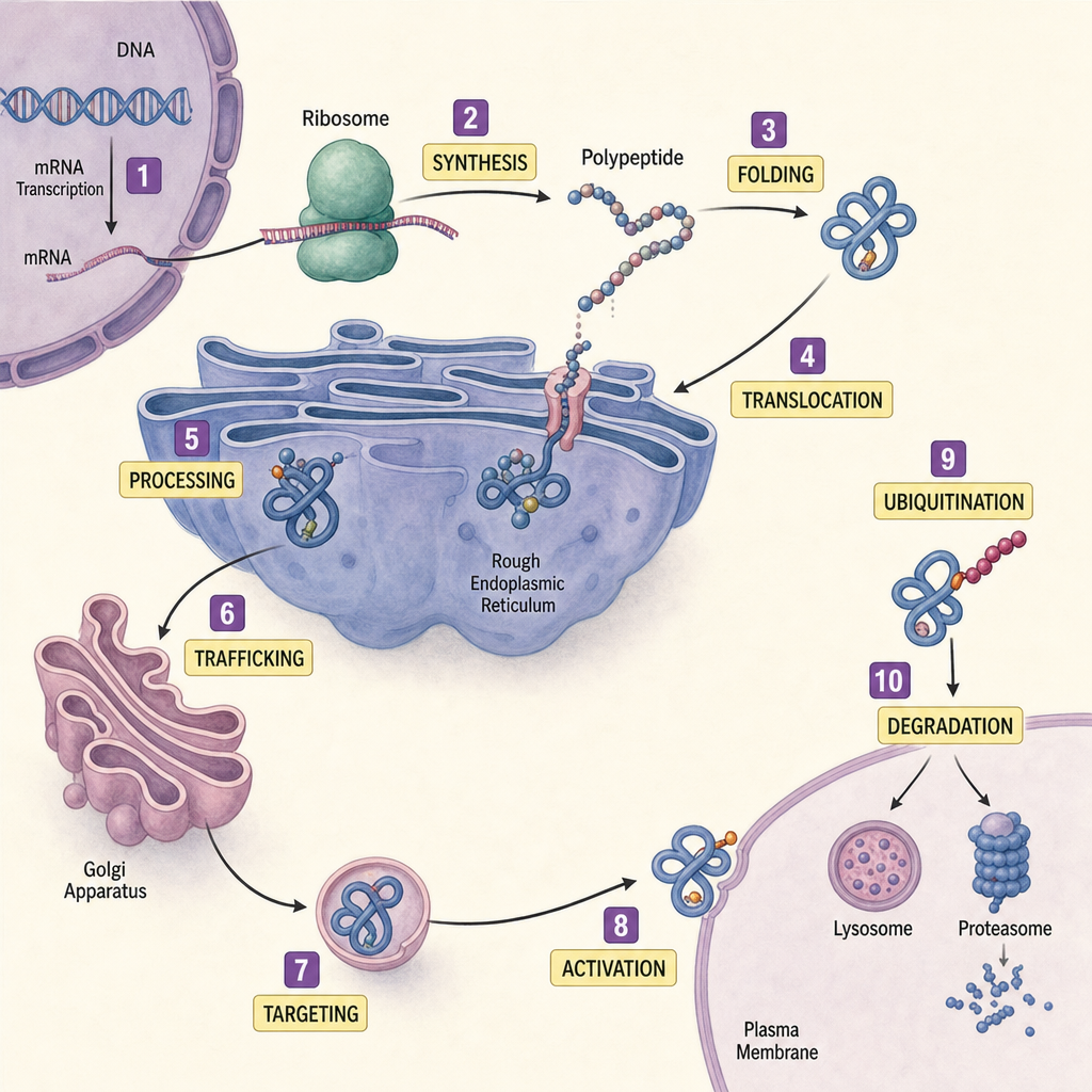
In the life cycle of a protein, what do stages 2, 5, and 9 represent?
Which of the following is an example of a conjugated protein?
Which of the following aids in the folding of proteins?
Which substance is commonly found in skin, hair, and nails?
Which of the following plays a central role in the mitotic spindle formation in cellular division?
Practice by Chapter
Amino Acids: Structure and Properties
Practice Questions
Peptide Bond Formation
Practice Questions
Primary Structure of Proteins
Practice Questions
Secondary Structure of Proteins
Practice Questions
Tertiary and Quaternary Structures
Practice Questions
Protein Folding and Chaperones
Practice Questions
Protein Domains and Motifs
Practice Questions
Structure-Function Relationships
Practice Questions
Hemoglobin and Myoglobin
Practice Questions
Collagen and Elastin
Practice Questions
Albumin and Plasma Proteins
Practice Questions
Post-Translational Modifications
Practice Questions
Want unlimited practice?
Get full access to all questions, explanations, and performance tracking.
Scan to download app