
Protein Structure and Function — MCQs

On this page
What is the primary function of the Golgi apparatus?
Thyroxine is transported by?
Which type of collagen is primarily found in bones?
Which of the following is the most abundant glycoprotein present in the basement membrane?
Which of the following represents the strongest type of interaction?
Which class of amino acids contains only non-essential amino acids?
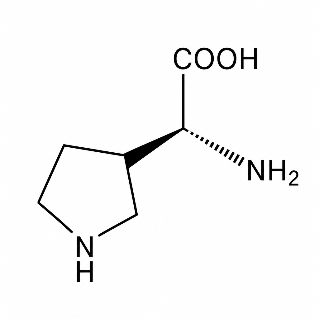
Which is the amino acid shown below?
Which of the following protein structures is not affected by denaturation?
Which of the following is a function of proteins containing a zinc finger motif?
Which type of glycoprotein linkage is found in collagen?
Practice by Chapter
Amino Acids: Structure and Properties
Practice Questions
Peptide Bond Formation
Practice Questions
Primary Structure of Proteins
Practice Questions
Secondary Structure of Proteins
Practice Questions
Tertiary and Quaternary Structures
Practice Questions
Protein Folding and Chaperones
Practice Questions
Protein Domains and Motifs
Practice Questions
Structure-Function Relationships
Practice Questions
Hemoglobin and Myoglobin
Practice Questions
Collagen and Elastin
Practice Questions
Albumin and Plasma Proteins
Practice Questions
Post-Translational Modifications
Practice Questions
Want unlimited practice?
Get full access to all questions, explanations, and performance tracking.
Scan to download app