
Protein Structure and Function — MCQs

On this page
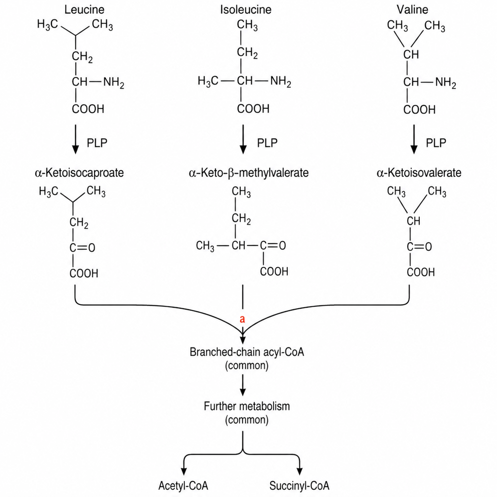
A 5-day-old neonate presents with poor feeding, progressive encephalopathy, and a sweet caramel-like odour to the urine. Plasma amino acid analysis shows markedly elevated leucine, isoleucine, and valine. The newborn screening card is flagged. The relevant catabolic pathway is shown (Image 1), depicting transamination of branched-chain amino acids (leucine, isoleucine, valine) to their corresponding branched-chain α-keto acids, followed by the shared oxidative decarboxylation step (marked with a bold red X) catalysed by the branched-chain α-keto acid dehydrogenase (BCKDH) complex, which converts the α-keto acids to their respective acyl-CoA products. Which coenzyme is directly required by the enzymatic complex blocked at the step indicated in the image?
Which of the following amino acids is not coded by a standard triplet codon?
Cathelicidins are rich in which of the following amino acids?
The conversion of an optically pure isomer (enantiomer) into a mixture of equal amounts of both dextro and levo forms is called as?
What are amphipathic helices?
Which of the following is NOT an intermediate filament?
All of the following are glycoproteins except?
Which of the following amino acid substitutions represents a conservative mutation?
Which of the following amino acids is a component of Thioredoxin reductase?
In which of the following is the highest concentration of cystine found?
Practice by Chapter
Amino Acids: Structure and Properties
Practice Questions
Peptide Bond Formation
Practice Questions
Primary Structure of Proteins
Practice Questions
Secondary Structure of Proteins
Practice Questions
Tertiary and Quaternary Structures
Practice Questions
Protein Folding and Chaperones
Practice Questions
Protein Domains and Motifs
Practice Questions
Structure-Function Relationships
Practice Questions
Hemoglobin and Myoglobin
Practice Questions
Collagen and Elastin
Practice Questions
Albumin and Plasma Proteins
Practice Questions
Post-Translational Modifications
Practice Questions
Want unlimited practice?
Get full access to all questions, explanations, and performance tracking.
Scan to download app