miRNA binds to which part of the mRNA to inhibit translation?
Steroid hormone receptors have attachment site for all except:
Which of the following statements regarding Huntington’s chorea is true?
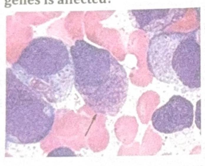
Identify the gene commonly involved in the condition shown in the image?

Which type of mutation can act as a suppressor to restore the wild-type phenotype in organisms carrying a mutant gene?
Which of the following is an X-linked dominant disorder?
Which one of the following is an autosomal dominant disorder?
Which one of the following statements about chromatin is not true?
Assertion: DNA methylation leads to gene silencing. Reason: Methylation prevents binding of transcription factors to promoter regions.
Phenotypic expression of a gene depending on the parent of origin is referred to as:
Explanation: ***3'UTR*** - MicroRNAs (miRNAs) are small non-coding RNA molecules that regulate gene expression. - They primarily bind to the **3' untranslated region (3'UTR)** of messenger RNA (mRNA) molecules, leading to translational repression or mRNA degradation. *Gene promoter* - The **gene promoter** is a region of DNA located upstream of a gene, where regulatory proteins bind to initiate transcription. - miRNAs do not directly bind to gene promoters to inhibit translation. *Gene body* - The **gene body** refers to the entire transcribed region of a gene, including exons and introns. - While some regulatory elements can be found within the gene body, the primary binding site for miRNAs to exert translational control is the 3'UTR. *5'UTR* - The **5' untranslated region (5'UTR)** is located at the 5' end of an mRNA molecule, upstream of the start codon. - While the 5'UTR can play a role in regulating translation initiation, it is not the primary target for miRNA binding to inhibit translation.
Explanation: ***Transcription activators*** - **Steroid hormone receptors** bind directly to **steroid hormones** and **hormone response elements (HREs)** on DNA, as well as to **transcription repressors** in their inactive state. - They do not have a direct attachment site for **transcription activators**; rather, activated steroid receptors can *act* as transcription activators or co-activators through protein-protein interactions. *Hormone responsive element* - This is a specific **DNA sequence** in the promoter region of target genes where the **steroid hormone-receptor complex** binds to regulate gene transcription. - It defines the genomic target for the activated steroid receptor, ensuring **gene-specific responses**. *Steroid hormone* - The **steroid hormone** itself binds to its specific receptor, inducing a conformational change that allows the receptor to translocate to the nucleus and bind to DNA. - This binding is essential for the **receptor's activation** and subsequent gene regulation. *Transcription repressors* - In the absence of a steroid hormone, **transcription repressors** (e.g., heat shock proteins) are often bound to the **steroid hormone receptor**, preventing its activation and binding to DNA. - Upon hormone binding, these repressors dissociate, allowing the receptor to become active and modulate **gene expression**.
Explanation: ***It is a trinucleotide repeat expansion type of disorder*** - Huntington's chorea is caused by an expansion of a **CAG trinucleotide repeat** in the **huntingtin gene (HTT)**. - This expansion leads to a misfolded protein and an **autosomal dominant** neurodegenerative disorder [1]. *There is a loss of function type of mutation.* - Huntington's chorea is primarily a **gain-of-function** mutation, where the expanded polyglutamine tract in the huntingtin protein leads to **toxic protein aggregation** and neuronal dysfunction. - While there might be some aspects of altered protein function, the core pathology is attributed to the **toxic effects** of the abnormal protein rather than a simple loss of its original function [1]. *It is an autosomal recessive disorder.* - Huntington's chorea is an **autosomal dominant** disorder, meaning only one copy of the mutated gene is sufficient to cause the disease. - Each child of an affected parent has a **50% chance** of inheriting the disease. *Increased number of CAA repeats.* - Huntington's chorea is characterized by an increased number of **CAG trinucleotide repeats**, not CAA repeats. - The expansion of these CAG repeats beyond a certain threshold (typically >35-40 repeats) in the huntingtin gene is directly responsible for the disease.
Explanation: ***BRAF V600E*** - The image displays cells with **Langerhans cell morphology**, including folded nuclei and abundant pale cytoplasm, which are characteristic of **Langerhans cell histiocytosis (LCH)** [1]. - The **BRAF V600E mutation** is the most common genetic alteration found in LCH, present in about 50-60% of cases and activating the MAPK pathway [1]. *RAS* - **RAS mutations** are frequently seen in various cancers, including colorectal adenocarcinoma, pancreatic adenocarcinoma, and non-small cell lung cancer. - While RAS pathway activation can occur in LCH, a direct RAS mutation is not the most common genetic driver; rather, downstream effectors like BRAF V600E are more prominent [1]. *RET* - **RET mutations** are primarily associated with **medullary thyroid carcinoma** (in both sporadic and inherited forms like MEN 2A and MEN 2B) and can also be found in certain types of lung cancer. - They are not a characteristic genetic alteration for Langerhans cell histiocytosis. *P53* - The **TP53 gene** encodes the tumor suppressor protein p53, and mutations in this gene are among the most frequent genetic alterations across a wide spectrum of human cancers. - Although p53 plays a critical role in cell cycle regulation and apoptosis, it is not a primary or common driver mutation specifically associated with Langerhans cell histiocytosis [1]. **References:** [1] Kumar V, Abbas AK, et al.. Robbins and Cotran Pathologic Basis of Disease. 9th ed. Diseases of White Blood Cells, Lymph Nodes, Spleen, and Thymus, pp. 629-630.
Explanation: ***Mutation of tRNA*** - A **tRNA suppressor mutation** can alter its anticodon, allowing it to recognize a **stop codon** (nonsense suppressor) or a missense codon, and insert an amino acid, thereby suppressing the original mutation. - This is a classic example of an **intergenic suppressor mutation** that acts at a different genetic locus from the original mutation. - These suppressors are particularly effective for **nonsense mutations** (premature stop codons) and certain missense mutations by correcting the decoding error during translation. *Frameshift mutation of coding gene* - A single frameshift mutation causes a shift in the **reading frame**, leading to a completely different protein sequence downstream and often a premature stop codon, which would worsen the phenotype. - While a **second compensating frameshift** mutation in the same gene could theoretically restore the reading frame (acting as an intragenic suppressor), this is context-dependent and less reliable than tRNA suppressors. - The question asks for mutations that "can act as a suppressor," and **tRNA mutations are the more universally recognized and reliable suppressor mechanism** in classical genetics. *Deletion of mutant gene* - **Deleting the mutant gene** removes the genetic information entirely but does not restore wild-type function; instead, it typically results in **loss of function** or complete absence of the protein. - This would lead to a **null phenotype** rather than restoration of wild-type phenotype, especially if the gene is essential. *Addition of another normal gene* - The **addition of another normal (wild-type) gene copy** provides a functional protein that can compensate for the mutant gene's deficiency. - While this can restore a wild-type phenotype, it represents **gene complementation** or gene therapy, not a true suppressor mutation that modifies the interpretation or expression of the existing mutant allele.
Explanation: ***Vitamin D resistant rickets*** - This is an **X-linked dominant** disorder characterized by impaired renal phosphate reabsorption and defective vitamin D metabolism, leading to **rickets-like symptoms** despite adequate vitamin D intake. - Affected individuals show **hypophosphatemia** and bone defects, with sons and daughters of affected fathers being affected. *Red-green color blindness* - This is an **X-linked recessive** disorder, meaning it primarily affects males and is passed from carrier mothers to their sons. - Affected individuals have difficulty distinguishing **red and green hues** due to defects in cone photoreceptors. *Achondroplasia* - This is an **autosomal dominant** disorder caused by a mutation in the **FGFR3 gene**, leading to dwarfism. - It is not X-linked and affects both sexes equally, with an affected individual having a 50% chance of passing it to each child. *Familial hypercholesterolemia* - This is an **autosomal dominant** metabolic disorder characterized by extremely high levels of **LDL cholesterol** due to defects in the LDL receptor. - It is not X-linked and can lead to premature cardiovascular disease, affecting both males and females.
Explanation: ***Hereditary spherocytosis*** - It is characterized by **autosomal dominant inheritance** [1], leading to the destruction of red blood cells. - Mutations in proteins that maintain the **red blood cell membrane** integrity result in spherocyte formation [1]. *Cystic fibrosis* - This condition follows a **autosomal recessive inheritance pattern**, requiring two copies of the mutated gene for disease manifestation. - It is caused by mutations in the **CFTR gene**, affecting chloride transport and leading to thick secretions. *G-6PD deficiency* - This disorder is inherited in an **X-linked recessive manner** [2], primarily affecting males and transmitted through carrier females. - Characterized by **hemolytic anemia** triggered by certain medications or infections, it does not follow dominant inheritance [2]. *Sickle cell anemia* - Sickle cell anemia is also an **autosomal recessive disorder** [3], meaning affected individuals must inherit two copies of the sickle cell gene. - It results in a mutation in the **HBB gene**, leading to the production of abnormal hemoglobin (HbS) [3].
Explanation: ***H2A-H2B bind to both the entry and exit ends of DNA in nucleosomes*** - This statement is **not entirely true** as presented because while **H2A-H2B dimers** do make contacts with DNA near entry/exit regions, they do not bind **exclusively** at these ends. - In the nucleosome structure, two H2A-H2B dimers flank the central **(H3-H4)₂ tetramer** and interact with DNA throughout approximately **30 base pairs on each side**. - The **entry and exit points** of nucleosomal DNA are primarily stabilized by **linker histones (H1)**, which bind to the dyad axis and linker DNA regions. - The statement oversimplifies the complex three-dimensional interactions within the nucleosome core particle. *DNA winds approximately 1.75 times around the nucleosomes* - This statement is **true**; approximately **1.65 to 1.75 turns** of DNA (about 146-147 base pairs) wrap around the **histone octamer** to form the core nucleosome particle. - This precise winding is crucial for the compaction of DNA into eukaryotic chromatin and represents the fundamental repeating unit of chromatin structure. *Covalent modification of histones influence chromatin compaction* - This statement is **true**; **post-translational modifications** (PTMs) such as acetylation, methylation, phosphorylation, and ubiquitination on histone tails significantly impact **chromatin structure and accessibility**. - For example, **histone acetylation** generally leads to a more open chromatin conformation (euchromatin) by neutralizing positive charges, facilitating gene expression. - **Histone methylation** can lead to either open or compact chromatin depending on the specific residue modified (e.g., H3K4me3 for activation, H3K9me3 for repression). *Non-histone proteins are part of mitotic chromosomes* - This statement is **true**; mitotic chromosomes contain numerous **non-histone proteins** essential for chromosome structure and function. - Examples include **structural maintenance of chromosomes (SMC) proteins** like condensin and cohesin, topoisomerases (DNA topoisomerase II), and kinetochore proteins. - These non-histone proteins are crucial for chromosome condensation, sister chromatid cohesion, segregation, and proper mitotic progression.
Explanation: ***Both assertion and reason are true and reason is the correct explanation.*** - **DNA methylation** at **CpG islands** in promoter regions is a well-established **epigenetic mechanism for gene silencing** - The reason directly explains HOW methylation causes silencing: **methylation prevents transcription factor binding** to promoter regions, blocking transcriptional machinery - Both statements are factually correct AND the reason provides the mechanistic explanation for the assertion *Assertion is true but reason is false.* - While the assertion is correct (DNA methylation does lead to gene silencing), the reason is also TRUE, not false - Methylation preventing transcription factor binding is indeed a **primary mechanism** of gene silencing - This option would only be correct if the reason statement were factually incorrect *Both assertion and reason are true but reason is not the correct explanation.* - Both statements are individually true, but this option is incorrect because the reason IS the correct explanation - The prevention of transcription factor binding **directly explains** how methylation silences genes - If this were correct, the reason would describe an unrelated consequence of methylation, not the causal mechanism *Both assertion and reason are false.* - Both statements are well-established biological facts - DNA methylation-mediated gene silencing is a fundamental epigenetic mechanism - Prevention of transcription factor binding is a validated mechanism of this silencing
Explanation: ***Genomic imprinting (parent-of-origin gene expression)*** - **Genomic imprinting** is an epigenetic phenomenon where gene expression is dependent on whether the gene was inherited from the mother or the father. - This results in monoallelic expression of specific genes, with only one copy (maternal or paternal) being active. *Mosaic genetic variation* - **Mosaicism** refers to the presence of two or more populations of genetically different cells in one individual, all derived from a single zygote. - This typically arises from a somatic mutation during development, not from differential expression based on parental origin. *Nonpenetrance of genotype* - **Nonpenetrance** occurs when individuals carrying a disease-causing genotype do not express the associated phenotype. - This concept relates to the presence or absence of a phenotype, not the differential expression based on parental origin. *Genetic anticipation* - **Genetic anticipation** is the phenomenon where the symptoms of a genetic disorder become more severe and/or appear at an earlier age in successive generations. - This is commonly observed in disorders caused by expansions of trinucleotide repeats, such as Huntington's disease, and is distinct from parent-of-origin gene expression.
Get full access to all questions, explanations, and performance tracking.
Start For Free