Activated protein C inhibits the clotting mechanism by inactivating which of the following clotting factors?
Von Willebrand disease involves a deficiency of which factor?
Which of the following statements is true regarding von Willebrand disease?
The clot formed after the coagulation cascade is not stable unless extensive cross-linking occurs. This is done by:
A patient on low-molecular-weight heparin suddenly develops a severe hemorrhage. What test would be most useful to assess the degree of anticoagulation?
Which of the following clotting factors in a patient on Warfarin therapy would show the earliest decrease in functional activity?
Which of the following is not typically seen in Disseminated Intravascular Coagulation (DIC)?
A 34-year-old, G1P0, presents for genetic counseling at 12 weeks' gestation. The patient has two sisters and a brother; her father has hemophilia. Her siblings are not affected, but she has a nephew who is affected. What is the inheritance pattern of this disorder?
Which of the following is a sex-linked disorder?
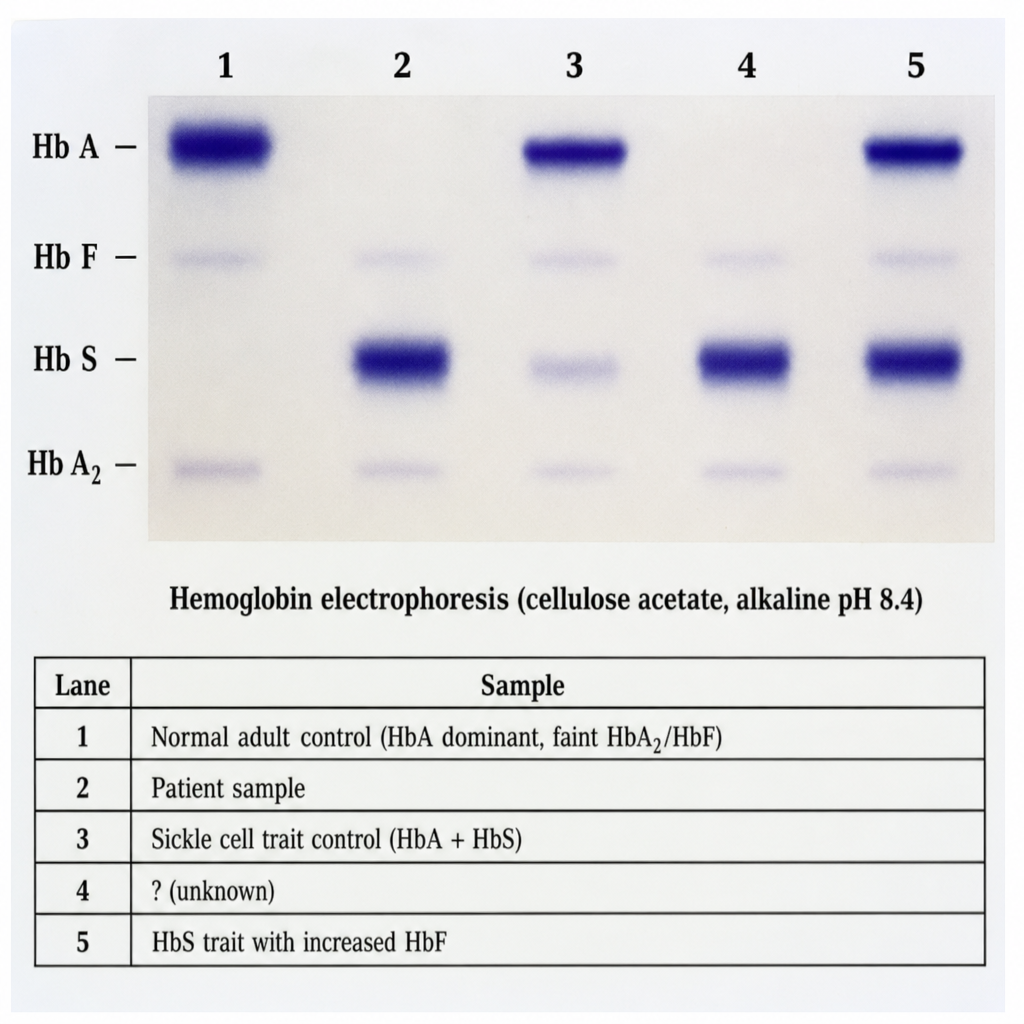
Ferritin biosynthesis is regulated by the serum level of which substance?
Explanation: ***Factor Va and Factor VIIIa*** - **Activated protein C (APC)** functions as a natural anticoagulant by specifically inactivating the activated forms of **Factor V (Va)** and **Factor VIII (VIIIa)**. - By inactivating these cofactors, APC effectively downregulates the functioning of the **prothrombinase complex** and **tenase complex**, thereby slowing down thrombin generation and subsequent fibrin formation. *Factor III and Factor VIIIa* - **Factor III (tissue factor)** initiates the extrinsic coagulation pathway, but it is not directly inactivated by activated protein C. - While **Factor VIIIa** is a target of APC, the combination with Factor III makes this option incorrect. *Factor VIIIa and Factor IX* - **Factor VIIIa** is indeed inactivated by APC. - However, **Factor IX** (the inactive zymogen) and its activated form **Factor IXa** are not direct targets for inactivation by APC; Factor IXa remains active to participate in the tenase complex until its co-factor Factor VIIIa is inactivated. *Factor Va and Factor VII* - **Factor Va** is a known target for inactivation by APC. - **Factor VII** (the inactive zymogen) and its activated form **Factor VIIa** are not inactivated by APC; Factor VIIa plays a role in the initiation of coagulation by forming a complex with tissue factor.
Explanation: ***Primary hemostasis*** - Von Willebrand disease primarily affects **primary hemostasis** due to defective or deficient **von Willebrand factor (vWF)**, which is crucial for platelet adhesion [1][3]. - This defect results in **increased bleeding tendencies**, exemplified by symptoms like easy bruising and prolonged bleeding from cuts [2]. *Secondary hemostasis* - Secondary hemostasis involves the **coagulation cascade**, not primarily affected in von Willebrand disease [3]. - Disorders related to secondary hemostasis typically involve factors like **factor VII, IX, or X**, unlike the vWF defect seen here [3]. *Generalized defects involving small vessels* - Generalized defects imply broader issues affecting the **microcirculation**, which is not the primary issue in von Willebrand disease. - While small vessel bleeding can occur, it is not specific to this condition as it does not primarily involve the **platelet aggregation** defect [1]. *Clot stabilization and resorption* - Clot stabilization and resorption primarily involve factors such as **fibrinogen** and cross-linking factors, rather than vWF. - Von Willebrand disease specifically impacts the **platelet function** and does not directly relate to stabilization processes once the clot has formed [1][3].
Explanation: Normal prothrombin time (PT) - In von Willebrand disease, **PT remains normal**, which indicates that the extrinsic pathway of coagulation is unaffected [1]. - This disorder primarily affects **platelet function** and vWF levels, not prothrombin time. *Platelet count may be decreased in some cases* - While platelet count can be low, it is not a consistent finding in von Willebrand disease; often, **platelet count is normal**. - The disorder primarily involves **qualitative abnormalities** in platelets due to impaired vWF function, rather than quantitative [3]. *Bleeding time is prolonged* - Bleeding time is typically **prolonged** in von Willebrand disease, which reflects platelet dysfunction, but this statement does not correctly state its context. - The disease affects **hemostasis**, leading to increased bleeding tendencies rather than maintaining normal bleeding times. *Normal activated partial thromboplastin time (aPTT)* - In von Willebrand disease, **aPTT may be prolonged** due to the deficiency of factor VIII, which is carried by vWF [2]. - The presence of normal aPTT does not reflect the disease's impact on the intrinsic pathway of coagulation.
Explanation: ***Factor XIII*** - **Factor XIIIa** (activated Factor XIII) is a **transglutaminase** that catalyzes the formation of **covalent bonds** between **fibrin monomers**, specifically between lysine and glutamine residues. - This cross-linking strengthens the **fibrin clot**, making it more resistant to mechanical stress and proteolytic degradation. *Plasmin* - **Plasmin** is an enzyme responsible for **fibrinolysis**, meaning it breaks down fibrin clots. - It acts to remodel and **dissolve clots**, not to stabilize them. *Thrombin* - **Thrombin** (Factor IIa) is a key enzyme in the coagulation cascade that converts **fibrinogen into fibrin monomers**. - While essential for clot formation, thrombin's primary role is to create the fibrin mesh, not to extensively cross-link it for stability. *High molecular weight kininogen* - **High molecular weight kininogen (HMWK)** is a cofactor in the **intrinsic coagulation pathway**, facilitating the activation of Factor XII and prekallikrein. - It is involved in initiating coagulation but does not directly participate in the cross-linking and stabilization of the fibrin clot.
Explanation: ***Stop LMWH immediately*** - The immediate priority in severe hemorrhage due to **low-molecular-weight heparin (LMWH)** is to cease further administration of the anticoagulant. [1] - This prevents worsening of the bleeding by halting the delivery of more drug that could contribute to the **anticoagulant effect**, thus allowing natural clotting mechanisms to begin recovery. [1] *Administer protamine sulfate* - While **protamine sulfate** is a reversal agent for LMWH, its efficacy is only partial (about 60-75% neutralization of anti-Xa activity) compared to its effect on unfractionated heparin. - Therefore, it is a secondary step after stopping the drug itself and may not fully reverse the severe hemorrhage. *Transfuse blood products* - **Transfusion of blood products** (e.g., packed red blood cells, fresh frozen plasma, platelets) addresses the consequences of severe hemorrhage (e.g., anemia, coagulopathy) but does not directly neutralize the anticoagulant effect of LMWH. - This is a supportive measure, crucial for managing blood loss and maintaining hemodynamic stability, but not the primary action to stop the drug's effect. *Consider specific reversal agents* - **Specific reversal agents** for LMWH itself are limited; protamine sulfate is the primary option, albeit with partial efficacy. - Newer agents for direct oral anticoagulants might be considered in other contexts, but for LMWH, stopping the drug is the most immediate and universally applicable action.
Explanation: ***Factor VII*** - Factor VII has the **shortest half-life** (approximately 6 hours) among the vitamin K-dependent clotting factors, meaning its functional activity decreases **most rapidly** after starting warfarin therapy. - Warfarin inhibits vitamin K epoxide reductase, preventing gamma-carboxylation of **all vitamin K-dependent factors** (II, VII, IX, X). However, Factor VII's short half-life means pre-existing functional Factor VII is depleted first. - This is why **PT/INR** (which measures the extrinsic pathway dependent on Factor VII) rises before aPTT in warfarin therapy. - Reduced gamma-carboxylation impairs Factor VII's ability to bind calcium and phospholipids, essential for its activation in the extrinsic coagulation pathway. *Factor IX* - Factor IX is a **vitamin K-dependent factor** affected by warfarin, but its longer half-life (approximately 24 hours) means functional activity decreases more slowly than Factor VII. - It plays a key role in the **intrinsic coagulation pathway**. *Factor X* - Factor X is a **vitamin K-dependent clotting factor** whose gamma-carboxylation is inhibited by warfarin. - Its half-life (approximately 40 hours) is longer than Factor VII, resulting in a **slower decline in functional activity**. *Prothrombin (Factor II)* - Prothrombin (Factor II) is a **vitamin K-dependent factor** affected by warfarin. - It has the **longest half-life** (60-72 hours) among vitamin K-dependent factors, meaning its functional levels decrease most slowly after initiating warfarin therapy.
Explanation: ***Normal APTT*** - In Disseminated Intravascular Coagulation (**DIC**), **APTT** is typically **prolonged** due to consumption of clotting factors [1]. - The presence of normal APTT indicates that coagulation pathways are not significantly affected, which is contrary to what is seen in DIC. *Fibrinogen decreased* - **Decreased fibrinogen levels** are common in DIC, reflecting its consumption during the coagulation process [1]. - This depletion is linked to the increased clotting and is a hallmark of DIC, making this statement false in the context of the question. *Thrombocytopenia* - **Thrombocytopenia** occurs in DIC as platelets are consumed during the formation of microclots [1]. - A significant drop in platelet count is a key feature of DIC, therefore this statement does not align with the "except" clause. *PT elevation* - Prothrombin Time (**PT**) is usually **elevated** in DIC due to the consumption of clotting factors [1]. - This reflects the ongoing activation of the coagulation cascade, supporting the exclusion in the question context. **References:** [1] Cross SS. Underwood's Pathology: A Clinical Approach. 6th ed. Common Clinical Problems From Blood And Bone Marrow Disease, pp. 625-626.
Explanation: ***X-linked inheritance*** - Hemophilia is an **X-linked recessive disorder** - An affected father passes his X chromosome mutation to **all daughters**, making them **obligate carriers** (not affected but carry the gene) - The affected nephew (son of patient's sister) confirms the patient's sister is a carrier who passed the affected X chromosome to her son - Classic pattern: affected males, carrier females, skips generations through female carriers *Autosomal recessive* - Would require both parents to be carriers for offspring to be affected - An affected father would pass one mutant allele to all children, but this wouldn't make daughters obligate carriers unless mother also carried the gene - Pattern of father → carrier daughter → affected grandson is not typical of autosomal recessive inheritance *Mitochondrial inheritance* - Only transmitted from mother to **all children** regardless of gender - Affected father **cannot** pass mitochondrial disorders to offspring - Would show maternal transmission pattern with all children of affected mothers being affected *Multifactorial inheritance* - Involves combination of multiple genes and environmental factors - Does not follow clear Mendelian pattern - The distinct single-gene pattern (affected father, carrier daughters, affected male grandchild) indicates X-linked recessive, not multifactorial
Explanation: ***Hemophilia*** - Hemophilia is an **X-linked recessive disorder**, meaning the gene responsible is located on the X chromosome. - Males are predominantly affected because they have only one X chromosome, so a single copy of the mutated gene is sufficient to cause the disease. *Neurofibromatosis* - Neurofibromatosis is an **autosomal dominant disorder**, meaning a single copy of the mutated gene on a non-sex chromosome is enough to cause the condition. - It affects males and females equally and is characterized by tumors along nerves and skin changes. *Klinefelter's syndrome* - Klinefelter's syndrome is a **chromosomal disorder** resulting from an extra X chromosome in males (XXY), not a single gene mutation on a sex chromosome. - While it involves sex chromosomes, it's categorized as a **sex chromosome aneuploidy** rather than a sex-linked disorder in the traditional genetic sense. *Thalassemia* - Thalassemia is an **autosomal recessive disorder**, meaning it requires two copies of the mutated gene (one from each parent) on non-sex chromosomes to manifest. - It affects the production of hemoglobin and impacts males and females equally.
Explanation: ### Explanation **Correct Option: C. Iron** Ferritin is the primary intracellular storage form of iron. Its biosynthesis is regulated at the **translational level** by the availability of free intracellular iron. This occurs via the **Iron Response Element (IRE)** and **Iron Regulatory Protein (IRP)** mechanism: * **Low Iron:** IRPs bind to the IRE located at the 5' untranslated region (UTR) of ferritin mRNA, physically blocking translation to prevent unnecessary storage. * **High Iron:** Iron binds to IRPs, causing them to dissociate from the mRNA. This allows the ribosome to translate the mRNA, increasing ferritin synthesis to safely sequester the excess iron. **Analysis of Incorrect Options:** * **A. Ceruloplasmin:** This is a copper-containing ferroxidase that converts Fe²⁺ to Fe³⁺ to facilitate iron binding to transferrin. It does not directly regulate ferritin synthesis. * **B. Hepcidin:** Known as the "Master Regulator of Iron Homeostasis," hepcidin controls systemic iron levels by degrading **ferroportin** (the iron exporter). While it influences iron availability, it does not directly regulate the biosynthesis of the ferritin protein. * **D. Transferrin:** This is the transport protein for iron in the plasma. While transferrin levels are inversely related to iron stores, it is a carrier, not a regulator of ferritin production. **High-Yield Clinical Pearls for NEET-PG:** * **Translational Control:** Ferritin is a classic example of post-transcriptional/translational regulation (unlike most proteins regulated at the transcriptional level). * **Serum Ferritin:** It is the **best initial test** and most sensitive marker for diagnosing **Iron Deficiency Anemia** (levels <15 ng/mL). * **Acute Phase Reactant:** Ferritin levels rise during inflammation, infection, or malignancy, which can mask an underlying iron deficiency. * **Hemosiderin:** This is an insoluble form of partially denatured ferritin, found in states of iron overload (e.g., Hemochromatosis).
Get full access to all questions, explanations, and performance tracking.
Start For Free