
Biochemical Techniques — MCQs

On this page
Which of the following statements is true regarding High-Performance Liquid Chromatography (HPLC) and Gas Chromatography?
Cells can be separated based on specific antigen receptors using which technique?
What is the method used to locate the isoelectric point of a protein?
If a radioimmunoassay is properly conducted and the amount of radioactive hormone bound to antibody is low, what would this result indicate?
Which assay is considered the most accurate for assessing hormone levels?
Method of chromatography in which molecules that are negatively charged are selectively released from the stationary phase by positively charged molecules in the mobile phase is termed as?
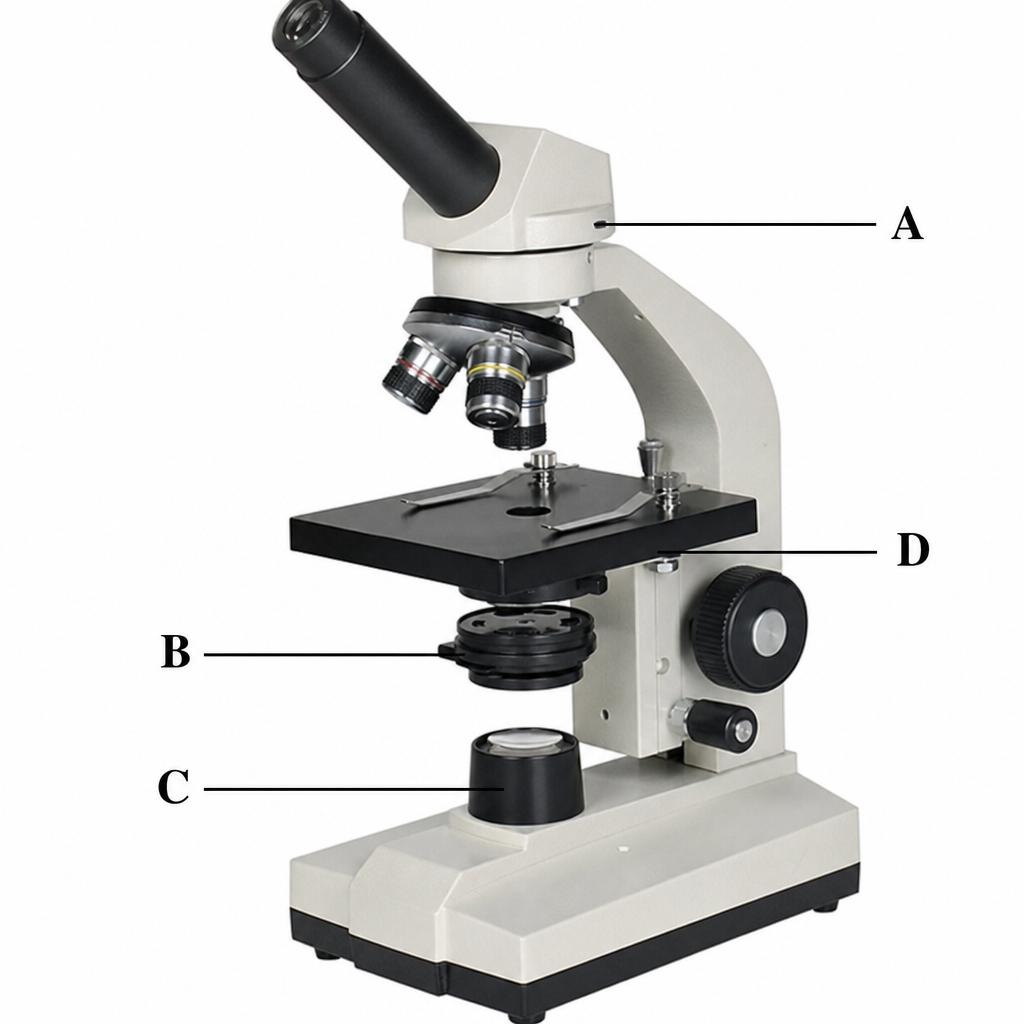
Which of the following labels corresponds to the condenser of the microscope?
Which of the following methods most accurately estimates blood creatinine level?
Protein is purified using ammonium sulfate by which method?
All of the following techniques can be used to determine protein structure, EXCEPT:
Practice by Chapter
Spectrophotometry and Colorimetry
Practice Questions
Chromatography Techniques
Practice Questions
Electrophoresis and Applications
Practice Questions
Centrifugation and Ultracentrifugation
Practice Questions
Radioisotope Techniques
Practice Questions
Enzyme-Linked Immunosorbent Assay (ELISA)
Practice Questions
Polymerase Chain Reaction (PCR)
Practice Questions
Blotting Techniques: Southern, Northern, Western
Practice Questions
Mass Spectrometry in Biochemistry
Practice Questions
Recombinant DNA Technology
Practice Questions
DNA Sequencing
Practice Questions
Proteomics and Metabolomics
Practice Questions
Want unlimited practice?
Get full access to all questions, explanations, and performance tracking.
Scan to download app