In PCR, DNA polymerase is used for which process?
What method is used to determine the tertiary structure of a protein?
Nephelometry is based on the principle of what?
Which amino acid migrates fastest on paper chromatography on methylcellulose medium?
Amplification of DNA uses the polymerase chain reaction (PCR) technique. Which cation is essential for PCR?
Match the following blotting techniques with the type of biomolecule they detect: Blotting Technique Detects A. Southern blot 1. RNA B. Northern blot 2. Lipids C. Western blot 3. DNA D. Eastern blot 4. Protein
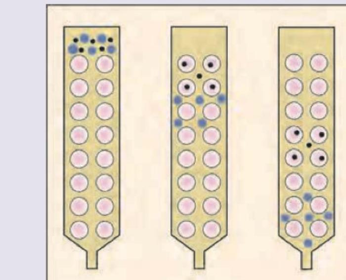
The following method of chromatography is called:

Which is the correct sequence of steps in isolating desirable protein using recombinant DNA technology? 1. Expression of protein and lysis of the bacterial cell 2. Incorporation of genes into bacteria 3. SDS PAGE 4. Protein elution 5. Column chromatography
Which of the following methods cannot be used to precipitate proteins?
Glycated hemoglobin (HbA1c) is best measured using?
Explanation: **Explanation:** **1. Why "DNA Multiplication" is the correct answer:** Polymerase Chain Reaction (PCR) is an *in vitro* technique used to generate millions of copies of a specific DNA segment from a minute starting sample. While the enzyme used (Taq Polymerase) performs the biochemical action of synthesis, the **ultimate objective and functional outcome** of using DNA polymerase in a PCR cycle is the exponential amplification or **multiplication** of the target genetic material. In the context of competitive exams like NEET-PG, when asked for the "process" PCR achieves via polymerase, "multiplication" (amplification) is the most comprehensive term describing the technique's purpose. **2. Analysis of Incorrect Options:** * **A. DNA Replication:** This is a biological process occurring *in vivo* (within a living cell) during the S-phase of the cell cycle. While PCR mimics this, it is a laboratory simulation, not the physiological process of replication. * **B. DNA Elongation:** This refers specifically to one sub-step of the PCR cycle (Extension). While the polymerase does elongate the primer, the question asks what the enzyme is used for in the context of the entire PCR process, which is to multiply the DNA. * **D. All of the above:** Since "Replication" is strictly a cellular term and "Elongation" is only a partial step, "Multiplication" stands as the most accurate descriptor for the technique's goal. **3. High-Yield Clinical Pearls for NEET-PG:** * **Taq Polymerase:** Derived from *Thermus aquaticus*; it is heat-stable, which is essential for the denaturation step (94-96°C). * **Steps of PCR:** Denaturation (95°C) → Annealing (55-65°C) → Extension (72°C). * **RT-PCR:** Used for RNA viruses (like SARS-CoV-2); involves Reverse Transcriptase to convert RNA to cDNA before amplification. * **Application:** PCR is the "gold standard" for diagnosing infectious diseases, genetic mutations, and forensic DNA profiling.
Explanation: ### Explanation **1. Why X-ray Crystallography is Correct:** X-ray crystallography is the gold-standard technique for determining the **three-dimensional (tertiary and quaternary) structure** of proteins at atomic resolution. The process involves growing a pure crystal of the protein and exposing it to an X-ray beam. The atoms in the crystal diffract the X-rays, creating a pattern that is mathematically analyzed to produce an **electron density map**. This allows scientists to map the precise spatial arrangement of amino acids, bonds, and secondary structures (alpha-helices and beta-sheets). **2. Why Other Options are Incorrect:** * **Spectrophotometry:** Used to measure the **concentration** of proteins or nucleic acids based on light absorption (e.g., proteins absorb UV light at 280 nm due to aromatic amino acids). It does not provide structural data. * **Electrophoresis (e.g., SDS-PAGE):** Used to separate proteins based on their **molecular weight** or charge. It tells us how large a protein is but not how it is folded. * **Chromatography:** A technique used for the **purification and separation** of protein mixtures based on size, charge, or affinity. **3. NEET-PG High-Yield Facts:** * **NMR Spectroscopy:** Another method for tertiary structure determination, specifically for proteins in **aqueous solution** (useful for proteins that won't crystallize). * **Cryo-Electron Microscopy (Cryo-EM):** An emerging high-yield topic; it is used for large macromolecular complexes that are difficult to crystallize. * **Ramachandran Plot:** Used to validate the secondary structure of proteins by plotting the dihedral angles (phi $\phi$ and psi $\psi$). * **Edman Degradation:** The classic method for determining the **primary sequence** (amino acid order) of a protein.
Explanation: **Explanation:** **Nephelometry** is a laboratory technique used to measure the concentration of substances (usually proteins) in a solution by measuring the **scattering of light** by suspended particles (antigen-antibody complexes). 1. **Why Option A is Correct:** When a beam of light passes through a solution containing suspended particles, the particles scatter the light in various directions. Nephelometry specifically measures the intensity of this **scattered light**, typically at an angle (usually 90°) to the incident beam. The intensity of the scattered light is directly proportional to the number of particles in the solution. Therefore, the light is "attenuated" (reduced/changed) in its original path due to the scattering effect. 2. **Why Other Options are Incorrect:** * **Option B (Refraction):** Refraction is the bending of light as it passes from one medium to another (e.g., used in refractometry to measure total serum protein). It is not the basis for nephelometry. * **Option C (Reduced transmission):** This describes **Turbidimetry**. While both involve light and particles, turbidimetry measures the *decrease* in the intensity of the light beam as it passes straight through the solution (180°), whereas nephelometry measures the light scattered at an angle. * **Option D (Filtration):** This refers to physiological renal clearance and has no relation to optical biochemical techniques. **High-Yield Clinical Pearls for NEET-PG:** * **Clinical Use:** Nephelometry is the "Gold Standard" for quantifying **Immunoglobulins (IgG, IgA, IgM)**, Complement proteins (C3, C4), and Acute Phase Reactants like **CRP**. * **Sensitivity:** Nephelometry is significantly **more sensitive** than turbidimetry for detecting low concentrations of proteins. * **Key Difference:** Remember, **N**ephelometry = **N**inety degrees (measures scattered light), **T**urbidimetry = **T**ransmission (measures unscattered light).
Explanation: ### Explanation The migration of amino acids in paper chromatography is primarily determined by their **partition coefficient** between the stationary phase (water bound to cellulose) and the mobile phase (organic solvent). **1. Why Valine is Correct:** Paper chromatography is a type of partition chromatography. The mobile phase is typically non-polar, while the stationary phase is polar. Amino acids with **non-polar (hydrophobic) side chains** have a higher affinity for the mobile phase and travel further and faster. **Valine**, being a branched-chain amino acid with a non-polar aliphatic side chain, is the most hydrophobic among the options provided. Therefore, it migrates the fastest and has the highest **$R_f$ (Retention factor) value**. **2. Analysis of Incorrect Options:** * **Aspartic Acid (Option A):** This is an acidic, highly polar amino acid. It has a high affinity for the aqueous stationary phase and thus migrates the slowest. * **Lysine (Option C):** This is a basic, positively charged polar amino acid. Like aspartic acid, its high polarity keeps it bound more tightly to the stationary phase, resulting in slow migration. * **Glycine (Option D):** While glycine is technically non-polar, its side chain is only a hydrogen atom. It is significantly less hydrophobic than valine and thus migrates slower than valine but faster than the charged amino acids. **3. NEET-PG High-Yield Pearls:** * **$R_f$ Value:** $R_f = \text{Distance traveled by solute} / \text{Distance traveled by solvent}$. It is always $\leq 1$. * **Hydrophobicity Trend:** In standard paper chromatography, the migration speed follows the order: **Non-polar > Uncharged Polar > Charged Polar**. * **Ninhydrin Reaction:** Most amino acids react with ninhydrin to give a **Ruhemann's purple** color; however, **Proline and Hydroxyproline** give a characteristic **yellow** color. * **Identification:** Amino acids are identified by comparing their $R_f$ values to known standards.
Explanation: **Explanation:** The Polymerase Chain Reaction (PCR) is a fundamental molecular technique used to amplify specific DNA sequences. The correct cation required for this process is **Magnesium ($Mg^{2+}$)**, usually added in the form of Magnesium Chloride ($MgCl_2$). **Why Magnesium is Essential:** 1. **Cofactor for DNA Polymerase:** $Mg^{2+}$ acts as an essential inorganic cofactor for *Taq* polymerase. It is required for the enzyme's catalytic activity. 2. **dNTP Binding:** Magnesium ions bind to the alpha-phosphate groups of deoxynucleotide triphosphates (dNTPs), facilitating the formation of the phosphodiester bond between the 3' OH group of the primer and the phosphate group of the incoming dNTP. 3. **Primer-Template Stability:** $Mg^{2+}$ ions help stabilize the negative charges on the phosphate backbone of the DNA, promoting the hybridization (annealing) of primers to the target DNA template. **Analysis of Incorrect Options:** * **Calcium (A):** While calcium is a vital signaling cation in the body, it is not a cofactor for DNA polymerases and can actually inhibit PCR at high concentrations. * **Lithium (B):** Lithium is primarily used in psychiatry (mood stabilizer) and has no functional role in DNA replication or PCR. * **Sodium (D):** Sodium ions ($Na^+$) are used in buffers to maintain ionic strength, but they cannot replace the divalent $Mg^{2+}$ required for the enzymatic catalysis of DNA synthesis. **High-Yield Facts for NEET-PG:** * **Optimization:** $Mg^{2+}$ concentration is critical. Too little results in low yield; too much can lead to non-specific amplification (mispriming). * **Chelation:** EDTA (a chelating agent) can inhibit PCR by sequestering $Mg^{2+}$ ions. * **Taq Polymerase:** Derived from the thermophilic bacterium *Thermus aquaticus*, it remains stable at high temperatures (denaturation step). * **Components of PCR:** Template DNA, Primers, dNTPs, *Taq* Polymerase, and $Mg^{2+}$ buffer.
Explanation: ***A-3, B-1, C-4, D-2*** - **Southern blot** is named after Edwin Southern and is the foundational technique used to detect specific **DNA** sequences. - **Northern blot** detects specific **RNA** sequences, while **Western blot** targets specific **Proteins** using antibodies. ***A-1, B-3, C-2, D-4*** - This option incorrectly matches Southern blot (A) with RNA (1) and Northern blot (B) with DNA (3). - The standard nomenclature links Southern with **DNA** and Northern with **RNA** due to their evolutionary development from the original technique. ***A-4, B-2, C-1, D-3*** - This option incorrectly assigns Southern blot (A) to Protein (4) and Northern blot (B) to Lipids (2). - Western blot (C) detects **Protein**, not RNA (1), as suggested here. ***A-2, B-4, C-3, D-1*** - This option incorrectly matches Southern blot (A) with Lipids (2) and Western blot (C) with DNA (3). - **Eastern blot** (D) is a technique designed to detect **post-translational modifications** (like lipids and carbohydrates) on proteins, making the D-2 match plausible, but the other matches are incorrect (i.e., **Southern blot** detects DNA).
Explanation: ***Sephadex chromatography*** - The image depicts a column filled with beads (pink circles) that act as a **porous stationary phase**. Smaller molecules (black dots) enter the pores, while larger molecules (blue dots) bypass them, eluting faster due to their inability to penetrate the beads. - This separation based on **molecular size** is the principle of **size-exclusion chromatography** (also known as gel filtration or Sephadex chromatography), where Sephadex is a common matrix material. *Ion exchange chromatography* - This method separates molecules based on their **net charge**, using a stationary phase with charged groups that bind to oppositely charged molecules. - The image does not show any interaction based on charge; instead, it illustrates differences in how molecules navigate around or through the beads. *Gas liquid chromatography* - This technique separates volatile compounds based on their **differential partitioning** between a mobile gas phase and a stationary liquid phase coated on a solid support. - The illustration shows a liquid-phase column separation, not a gaseous mobile phase or vaporization of analytes. *Affinity chromatography* - This method separates molecules based on their **specific, reversible binding** to a ligand immobilized on the stationary phase. - The image does not indicate any specific binding interactions between the molecules being separated and the column matrix; rather, it shows physical exclusion based on size.
Explanation: ***2,1,3,5,4*** - This sequence accurately reflects the typical order of operations in **recombinant protein isolation**: first, the gene is introduced into bacteria, then protein is expressed and cells lysed, followed by **SDS-PAGE as an intermediate quality check** to confirm protein expression before proceeding to purification steps (column chromatography and elution). - The process starts with gene incorporation, includes an analytical checkpoint after lysis, and ends with purified protein elution. *2,4,5,3,1* - This sequence is incorrect because **protein elution (4)** and **column chromatography (5)** are purification steps that occur *after* protein expression and cell lysis. - **Lysis (1)** cannot happen after elution, as cells must be lysed first to release the protein for purification. *1,2,4,3,5* - This sequence is incorrect because **expression and lysis (1)** must occur *after* the gene has been **incorporated into bacteria (2)** - the gene must be present before it can be expressed. - Additionally, **protein elution (4)** should follow **column chromatography (5)**, as elution is the step where protein is collected from the chromatography column. *1,5,2,4,3* - This sequence is incorrect because **incorporation of genes (2)** must be the first step - the gene needs to be in the bacteria before any expression, lysis, or purification can occur. - Starting with **expression and lysis (1)** before gene incorporation is impossible.
Explanation: ***Moving pH away from isoelectric pH*** - Proteins are **least soluble** at their **isoelectric point (pI)**, where their net charge is zero, causing them to aggregate and precipitate. - Moving the pH **away from the isoelectric point** increases the net charge on the protein, enhancing its solubility and preventing precipitation. *Add alcohol and acetone* - **Organic solvents** like alcohol and acetone reduce the dielectric constant of water, weakening the **hydrophobic interactions** that maintain protein solubility. - This leads to increased protein-protein interactions and **precipitation** as the protein unfolds or aggregates. *Using heavy metal ions* - **Heavy metal ions** (e.g., lead, mercury) are positively charged and bind strongly to the negatively charged groups on proteins, such as **carboxylates** and **sulfhydryl groups**. - This binding can disrupt protein structure, lead to aggregation, and cause **precipitation**. *Adding trichloroacetic acid* - **Trichloroacetic acid (TCA)** is a strong acid that significantly lowers the pH of the solution, causing proteins to become **protonated**. - This change in charge and the disruption of **salt bridges** and hydrogen bonds lead to protein denaturation and **precipitation**.
Explanation: ***Ion exchange chromatography*** - This method separates hemoglobin variants based on their **charge differences** due to the glucose molecule attached to HbA1c. - It is a highly sensitive and specific method for quantifying HbA1c, widely used in clinical laboratories. *Isoelectric focusing* - This technique separates molecules based on their **isoelectric point (pI)**, the pH at which they have no net charge. - While it can differentiate some hemoglobin variants, it is generally **less efficient and more complex** for routine HbA1c measurement compared to ion exchange chromatography. *Affinity chromatography* - This method separates molecules based on their **specific binding affinity** to a ligand immobilized on a stationary phase. - While it has been explored for HbA1c measurement, it is **not the most commonly used** or preferred method due to potential interferences and cost compared to ion exchange chromatography. *Electrophoresis* - This technique separates molecules based on their **charge and size** in an electric field. - While it can separate major hemoglobin variants, it has **lower resolution and accuracy** for routine HbA1c quantification compared to more specialized chromatographic methods, making it less ideal for precise measurement.
Spectrophotometry and Colorimetry
Practice Questions
Chromatography Techniques
Practice Questions
Electrophoresis and Applications
Practice Questions
Centrifugation and Ultracentrifugation
Practice Questions
Radioisotope Techniques
Practice Questions
Enzyme-Linked Immunosorbent Assay (ELISA)
Practice Questions
Polymerase Chain Reaction (PCR)
Practice Questions
Blotting Techniques: Southern, Northern, Western
Practice Questions
Mass Spectrometry in Biochemistry
Practice Questions
Recombinant DNA Technology
Practice Questions
DNA Sequencing
Practice Questions
Proteomics and Metabolomics
Practice Questions
Get full access to all questions, explanations, and performance tracking.
Start For Free