A Patient presented to emergency with multiple rib fractures. He is conscious speaking single words. On examination, respiratory rate was 40/minute and BP was 90/40 mmHg. What is immediate next step?
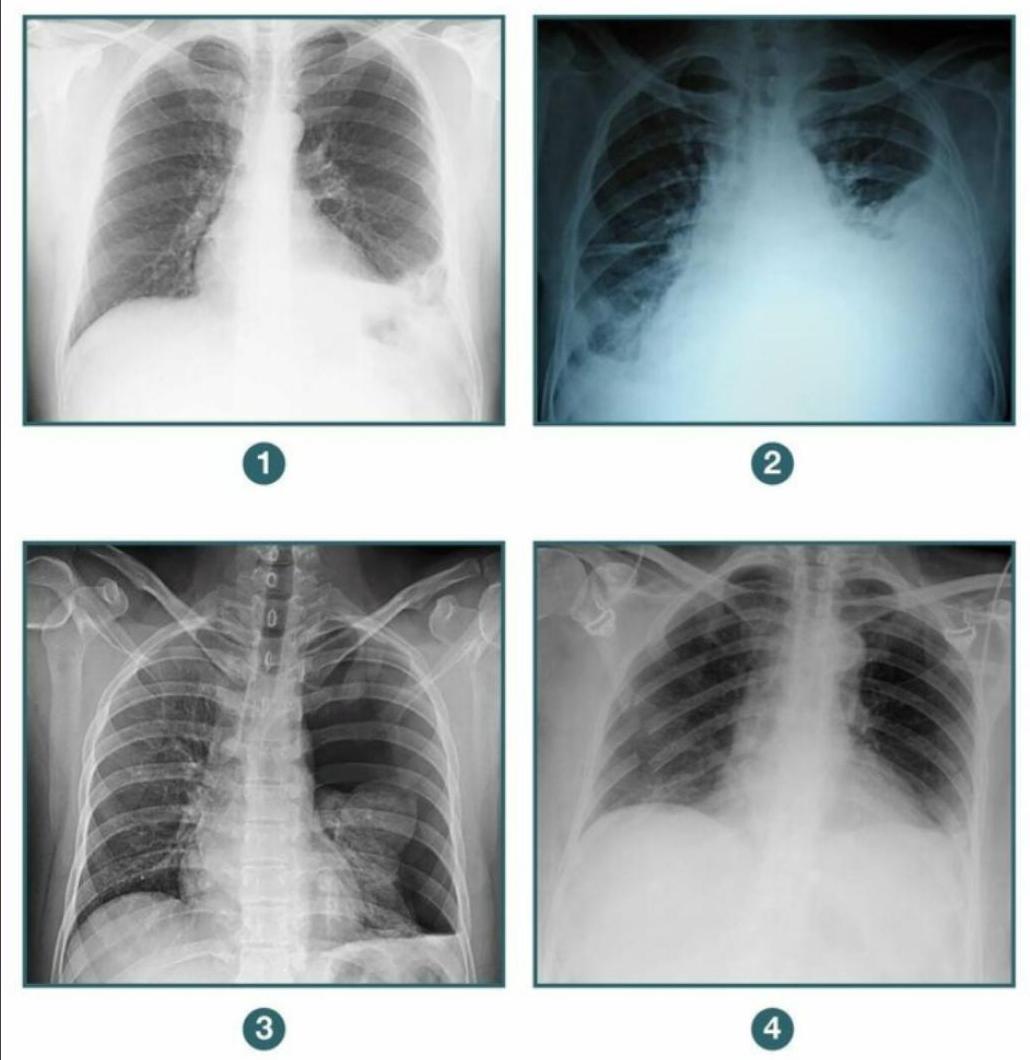
A patient presents to the casualty following blunt trauma to the chest. A chest X-ray was done. Among the following radiographs, in which case would you further evaluate the patient before putting a chest tube? 1. Diaphragmatic hernia 2. Hemothorax 3. Pneumothorax 4. Flail chest

Road traffic accident (RTA) with multiple fractures - initial treatment would be:
Indications for emergency thoracotomy are all of the following except:
A patient involved in a Road Traffic Accident (RTA) presents with: - Absent air entry on the left side of the chest. - Tenderness in the left lower chest wall. What is the next step in the Emergency Medicine Room (EMR) management?
What is to be addressed first in case of polytrauma -
A patient is admitted following a road traffic accident. He has sustained significant blunt injury to his head, chest and abdomen and has a Glasgow Coma Scale score of 8/15. His saturations are poor at 89% on 15 L of oxygen a rebreathing mask. You note bruising around both eyes and blood-stained fluid issuing from his left ear, which forms concentric circles when dripped on a white sheet. You wish to support his airway to improve oxygenation. The first choice of airway adjunct would be
Endotracheal tube in the esophagus is best assessed by:
Which of the following is an ideal method to prevent aspiration pneumonia?
Which of the following parameters is most critical for maintaining optimal oxygenation?
Explanation: ***Needle insertion in 2nd intercostal space*** - The patient's presentation with multiple rib fractures, **tachypnea (40/minute)**, **hypotension (90/40 mmHg)**, and speaking only single words suggests **tension pneumothorax**. - Speaking only single words indicates severe **respiratory distress** and inability to complete sentences due to dyspnea. - **Needle decompression** in the 2nd intercostal space at the midclavicular line is the immediate life-saving intervention for tension pneumothorax. - This is a **clinical diagnosis** in an emergency setting and does not require imaging confirmation before intervention. *Urgent IV fluid administration* - While fluid administration may be necessary for shock, the primary issue is likely **impaired ventilation** due to tension pneumothorax, which needs to be addressed first. - Delaying needle decompression to administer fluids could worsen the patient's respiratory and hemodynamic status. *Intubate the patient* - Intubation without addressing the cause of respiratory compromise, especially tension pneumothorax, can worsen the condition by increasing **intrathoracic pressure**. - Positive pressure ventilation in the presence of tension pneumothorax can be **life-threatening**. - **Relief of the tension pneumothorax** is the priority before considering definitive airway management. *Chest X-ray* - A chest X-ray is a diagnostic tool but should **not delay immediate life-saving interventions** in a patient with suspected tension pneumothorax. - Tension pneumothorax is a **clinical diagnosis** based on symptoms and immediate intervention takes precedence over imaging.
Explanation: ***Correct Option: Diaphragmatic hernia*** - A **diaphragmatic hernia** (showing elevated hemidiaphragm with loops of bowel in the hemithorax) requires **further evaluation before chest tube placement** - **CT scan with contrast** or **nasogastric tube with X-ray** should be performed to delineate the anatomy and confirm herniated abdominal contents - **Chest tube placement is contraindicated** or requires extreme caution as it could perforate herniated abdominal organs (stomach, bowel, liver, spleen) - This condition requires **surgical repair**, not chest drainage - The key principle: **Always evaluate thoroughly before intervention when diaphragmatic injury is suspected** *Incorrect Option: Pneumothorax* - A **pneumothorax** (characterized by absence of lung markings in the periphery and visceral pleural line) has a straightforward indication for chest tube - **Chest tube is the definitive management** for significant or symptomatic pneumothorax to re-expand the lung - No additional evaluation needed before chest tube placement in hemodynamically stable patients with confirmed pneumothorax *Incorrect Option: Hemothorax* - A **hemothorax** (showing opacification in the lower lung field with blunting of costophrenic angle and fluid level) has a clear indication for chest tube - **Chest tube is indicated** to drain blood, relieve lung compression, and monitor for ongoing bleeding - Immediate chest tube placement is appropriate once diagnosed *Incorrect Option: Flail chest* - A **flail chest** (multiple rib fractures in two or more places creating unstable chest wall segment) primarily requires **pain management and ventilatory support** - A chest tube is **not indicated for flail chest itself** unless there is an associated pneumothorax or hemothorax - If flail chest is isolated, you would not place a chest tube at all, making this option incorrect for the question asked
Explanation: ***Airway management*** - In trauma, **establishing and maintaining a patent airway** is the absolute priority, as compromised breathing can lead to rapid deterioration and death. - The **ABCs (Airway, Breathing, Circulation)** of trauma care dictate that airway intervention precedes other life-saving measures. *Management of shock* - While crucial, **managing shock (C)** follows **airway (A)** and **breathing (B)** in the primary survey of trauma care. - Addressing profound shock without a patent airway can be ineffective and leads to irreversible damage. *Splinting of limbs* - **Splinting fractures** is important for pain control, preventing further injury, and minimizing blood loss in open fractures, but it is not an immediate life-saving intervention. - This falls under the **secondary survey** or definitive management, after life-threatening issues have been addressed. *Cervical spine protection* - **Cervical spine protection** is essential in trauma to prevent further neurological injury and is performed simultaneously with airway management (often with in-line stabilization). - However, a patent airway is the **most immediate life-sustaining intervention** if the airway is compromised.
Explanation: ***Tension pneumothorax*** - While a life-threatening condition, a **tension pneumothorax** is initially managed with **needle decompression** or **chest tube insertion**, not an immediate emergency thoracotomy. - Emergency thoracotomy is reserved for situations requiring direct repair or control of massive bleeding that cannot be addressed by less invasive means. *Major tracheobronchial injuries* - These injuries can lead to severe **airway obstruction**, **massive air leak**, and **hemorrhage**, necessitating direct surgical repair via emergency thoracotomy. - Prompt surgical intervention is crucial to restore airway integrity and prevent life-threatening respiratory collapse. *Cardiac tamponade* - **Cardiac tamponade** can be caused by penetrating or blunt trauma, leading to circulatory collapse due to compression of the heart. - While initial management may involve pericardiocentesis, persistent or rapidly recurring tamponade, especially after trauma, often requires an **emergency thoracotomy** for direct repair of cardiac injury and evacuation of blood. *Penetrating injuries to anterior chest* - **Penetrating anterior chest injuries** carry a high risk of damage to vital structures such as the heart, great vessels, and major airways. - These injuries often result in rapid **hemodynamic instability**, severe hemorrhage, or cardiac arrest, making emergency thoracotomy essential for direct exploration and definitive repair.
Explanation: ***X-ray*** - In a **hemodynamically stable** patient with absent air entry and chest wall tenderness post-RTA, a **chest X-ray** is the most appropriate initial imaging in the EMR. - It quickly diagnoses conditions like **pneumothorax**, **hemothorax**, or **rib fractures** and guides management decisions. - **Important**: Clinical assessment for **tension pneumothorax** (hypotension, tracheal deviation, distended neck veins) must be done first. If tension pneumothorax is suspected, **immediate needle decompression** is required without waiting for imaging. - X-ray is **rapidly available** and provides crucial information for trauma management in stable patients. *FAST* - **Focused Assessment with Sonography for Trauma (FAST)** is primarily used to detect **intra-abdominal free fluid** (hemoperitoneum) or pericardial effusion in trauma. - While valuable in RTA evaluation, it is not the primary diagnostic tool for absent air entry in the chest. - FAST has limited sensitivity for **pneumothorax** and does not visualize **rib fractures** in detail. *DPL* - **Diagnostic Peritoneal Lavage (DPL)** is an invasive procedure used to detect **intra-abdominal injury** and hemorrhage. - It has largely been replaced by FAST and CT scans due to its invasive nature and lower specificity. - DPL provides **no information about chest injuries** and is irrelevant for evaluating absent air entry. *CT* - A **CT scan** (chest CT) provides highly detailed imaging and is excellent for diagnosing specific chest injuries. - However, it is **time-consuming**, requires patient transport, and is typically reserved for **stable patients** after initial X-ray assessment. - In the immediate EMR setting, X-ray is preferred for rapid decision-making, with CT used for further evaluation if needed.
Explanation: ***Airway*** - Maintaining a **patent airway** is the absolute first priority in polytrauma management according to the **ATLS (Advanced Trauma Life Support)** protocol. - Failure to secure an airway can lead to **hypoxia** and **brain damage** within minutes, regardless of other injuries. *Circulation* - While critical, addressing **circulation** (C in ABCDE) comes after establishing a secure airway and adequate breathing (A and B). - Uncontrolled hemorrhage would be the focus of circulation management, but only after guaranteeing proper oxygenation. *Neurology* - Neurological assessment (D in ABCDE for Disability) follows the primary survey of airway, breathing, and circulation. - Initial neurological evaluation focuses on **level of consciousness** using the **GCS (Glasgow Coma Scale)**. *Blood Pressure* - **Blood pressure** is an indicator of circulatory status but is not the first thing to be addressed. - It falls under the "C" for circulation in the ATLS protocol, which is secondary to airway and breathing.
Explanation: ***Oropharyngeal airway*** - An **oropharyngeal airway (OPA)** is the most appropriate initial airway adjunct in a patient with a **depressed GCS (8/15)** and poor oxygenation, as it helps to relieve **upper airway obstruction** caused by the tongue falling back. - Given the potential for a **basal skull fracture** (bruising around eyes, blood-stained fluid from ear forming concentric circles), a **nasopharyngeal airway (NPA)** is contraindicated due to the risk of intracranial insertion. *Nasopharyngeal tube* - A **nasopharyngeal airway (NPA)** is contraindicated in this patient due to signs suggestive of a **basal skull fracture**, which include **raccoon eyes (periorbital bruising)** and **Battle's sign (bruising behind the ear)**, as well as the **halo sign (concentric circles of blood and CSF)** from the ear. - Inserting an NPA in such a scenario risks inadvertently entering the **cranial cavity**, leading to further neurological damage or infection. *Intubation* - While **intubation** may eventually be necessary given the patient's low GCS and poor oxygenation, it is not the *first choice* of airway adjunct. - The immediate priority is to establish a **patent airway** quickly and safely, which an OPA can achieve while preparations for definitive intubation are made. *Laryngeal mask* - A **laryngeal mask airway (LMA)** could be considered for airway management, but it is typically a more advanced adjunct than an OPA. - Its insertion requires a certain level of skill and might be more time-consuming than an OPA, which is crucial in an emergency setting.
Explanation: ***CO2 Exhalation*** - Measuring **CO2 exhalation** (capnography) is the most reliable method to confirm endotracheal tube placement, as CO2 is present in the trachea but not in the esophagus. - A persistent **waveform on the capnograph** indicates proper tracheal intubation. *Direct laryngoscopy* - While helpful for initial visualization during intubation, **direct laryngoscopy** cannot confirm continuous tracheal placement after the tube is advanced. - It only confirms the tube passing through the vocal cords, not its final position in the trachea versus esophagus. *Auscultation* - **Auscultation** can be misleading because stomach sounds can be transmitted to the chest, and breath sounds can be heard in the epigastrium even with esophageal intubation. - It relies on subjective interpretation and is less definitive than capnography. *Chest wall movement* - Observing **chest wall movement** is not a definitive sign, as the chest can still rise with esophageal intubation due to air entering the stomach. - This method is unreliable and can be mistaken for proper ventilation, leading to dangerous delays in correcting tube misplacement.
Explanation: ***Endotracheal tube (cuffed)*** - A cuffed endotracheal tube forms a **seal** in the trachea, effectively preventing aspiration of gastric contents or oral secretions into the lungs. - This method is particularly crucial before and during surgical procedures involving general anesthesia, where normal airway protective reflexes are abolished. *Full stomach* - A **full stomach** significantly increases the risk of aspiration, as there is more gastric content available to be regurgitated into the airway. - This is a contraindication for immediate induction of general anesthesia and often necessitates a rapid sequence intubation. *Increase the intra abdominal pressure* - Increasing **intra-abdominal pressure** (e.g., due to obesity, insufflation for laparoscopy) can push gastric contents towards the esophagus, thereby increasing the risk of reflux and aspiration. - This effect is undesirable and directly contributes to aspiration risk rather than preventing it. *Inhalational anesthetic* - **Inhalational anesthetics** depress airway reflexes, making the patient more susceptible to aspiration. - While they are essential for maintaining anesthesia, they do not prevent aspiration; rather, other measures like intubation are necessary to counteract their effects.
Explanation: ***PEEP*** - **Positive End-Expiratory Pressure (PEEP)** is crucial for maintaining optimal oxygenation because it prevents **alveolar collapse** at the end of expiration, thereby increasing the **functional residual capacity** and improving gas exchange. - By keeping alveoli open, PEEP increases the number of available alveoli for ventilation, preventing **atelectasis** and optimizing the **venous admixture** from non-ventilated lung units. *FiO2* - While **Fraction of Inspired Oxygen (FiO2)** is essential for providing sufficient oxygen, simply increasing FiO2 without proper alveolar recruitment and patency (often achieved with PEEP) can be less effective and potentially harmful due to **oxygen toxicity**. - High FiO2 can improve oxygenation in cases of **hypoxemia**, but it doesn't address underlying problems like **alveolar collapse** or **ventilation-perfusion mismatch** as directly as PEEP does. *Respiratory rate* - **Respiratory rate** primarily affects **carbon dioxide elimination** (PaCO2) and, to some extent, alveolar ventilation. - While an adequate respiratory rate is necessary for overall gas exchange, it is not the most direct or critical parameter for optimizing **oxygenation** compared to PEEP's role in maintaining alveolar patency. *Tidal volume* - **Tidal volume** also primarily affects **carbon dioxide elimination** and plays a role in overall minute ventilation. - Excessive tidal volume can lead to **ventilator-induced lung injury (VILI)**, while insufficient tidal volume can reduce minute ventilation, but it does not directly optimize oxygenation by preventing **alveolar collapse** in the same way PEEP does.
Get full access to all questions, explanations, and performance tracking.
Start For Free