Which of the following surgical incisions is associated with the highest risk of postoperative pulmonary complications ?
What happens to gas exchange when the Va/Q ratio approaches infinity?
At the end of anaesthesia after discontinuation of nitrous oxide and removal of endotracheal tube, 100% oxygen is administered to the patient to prevent:
Procedure of choice for control of massive hemoptysis?
Which of the following is not true about ventilation-perfusion ratio (V/Q)?
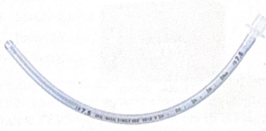
Identify the instrument shown in the image:

A man is brought to casualty who met with an accident. He sustained multiple rib fractures with paradoxical movement of chest. Management is:
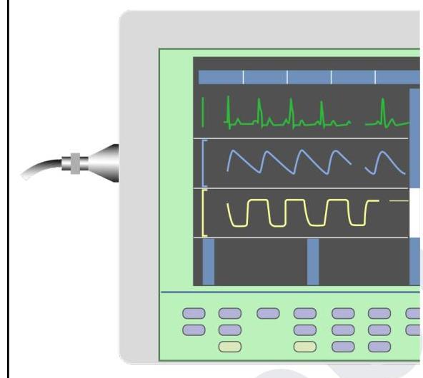
You are in the operating room and notice the tracing in yellow colour on this device. What does it indicate?

A 10-year-old boy, unconscious with 2 days history of fever, comes to pediatric ICU with respiratory rate 46/min, blood pressure 110/80 mmHg, and Glasgow Coma Scale E1 V1 M3. The next step in management is
A 30-year-old male was intubated for surgery. What is the best method to confirm the correct position of the endotracheal tube?
Explanation: ***Lateral thoracotomy*** - **Lateral thoracotomy** is associated with the **highest risk of postoperative pulmonary complications** among common surgical incisions, with complication rates ranging from **15-70%** depending on the procedure. - This incision **directly violates the chest wall** with rib resection or spreading, causing severe postoperative pain that significantly impairs respiratory mechanics. - The procedure disrupts **intercostal muscles**, damages **intercostal nerves**, and violates the **pleura**, leading to immediate risks like **pneumothorax**, **hemothorax**, and **pleural effusion**. - Severe pain leads to **splinting**, **shallow breathing**, **impaired cough**, and **reduced lung expansion**, markedly increasing the risk of **atelectasis**, **pneumonia**, and **respiratory failure**. - The **ipsilateral lung** is particularly affected with reduced functional residual capacity and impaired secretion clearance. *Vertical laparotomy* - **Upper abdominal vertical incisions** are indeed associated with high pulmonary complication rates (**30-50%**), second only to thoracotomy. - Pain leads to **diaphragmatic splinting** and impaired respiratory mechanics, increasing risk of **atelectasis** and **pneumonia**. - However, the chest wall itself remains intact, making complications generally less severe than with thoracotomy. *Median sternotomy* - While a major thoracic procedure, **median sternotomy** has relatively **lower pulmonary complication rates** compared to lateral thoracotomy. - The sternal split preserves **intercostal muscles** and **nerve integrity**, resulting in less severe pain and better preserved respiratory mechanics. - Postoperative pain management is generally more effective than with lateral thoracotomy. *Horizontal laparotomy* - **Transverse abdominal incisions** (e.g., Pfannenstiel, transverse supraumbilical) cause significantly less pain than vertical incisions. - These incisions follow **natural tissue planes**, cause less muscle disruption, and allow better respiratory mechanics. - Lower pain levels facilitate **effective coughing**, **deep breathing**, and **early mobilization**, reducing pulmonary complication risk.
Explanation: ***No exchange of O2 and CO2 occurs.*** - When the **Va/Q ratio approaches infinity**, it signifies a scenario of **ventilation without perfusion** (Q approaches zero). - This represents **alveolar dead space** - despite adequate ventilation, there is **no blood flow** to participate in gas exchange. - Therefore, **no O2 enters the blood** and **no CO2 leaves the blood**, making this the most accurate description of what happens to gas exchange. *Partial pressure of O2 becomes negligible.* - This statement is incorrect because with **no blood flow** (Q = 0), the alveolar air retains high O2 partial pressure. - O2 is being delivered via ventilation but not removed by blood, so **alveolar PO2** would approach that of **inspired air (~150 mmHg)**, not become negligible. *Partial pressure of CO2 becomes negligible.* - While this statement is technically true (alveolar PCO2 would approach zero/inspired air levels), it doesn't directly answer what happens to **gas exchange**. - With no blood flowing through the alveolus, no **CO2 from venous blood** can reach the alveolus to be excreted. - However, the question asks about **gas exchange** itself, not just partial pressures, making the first option more comprehensive. *Partial pressures of both CO2 and O2 remain normal.* - This statement is incorrect as the **Va/Q mismatch** significantly alters the partial pressures of both gases. - In infinite Va/Q scenario (dead space ventilation), **alveolar PO2 would be high** (approaching inspired air ~150 mmHg) and **alveolar PCO2 would be low** (approaching zero).
Explanation: ***Diffusion Hypoxia*** - Post-anaesthesia administration of 100% oxygen prevents **diffusion hypoxia**, a phenomenon where **nitrous oxide** rapidly diffuses out of the blood into the alveoli, diluting alveolar oxygen and carbon dioxide. - This rapid outflow of nitrous oxide can lead to a significant drop in **partial pressure of oxygen** in the alveoli, causing hypoxemia if not counteracted with high inspired oxygen. *Second gas effect* - The **second gas effect** refers to the phenomenon where the rapid uptake of a highly soluble anesthetic (like nitrous oxide) accelerates the uptake of a co-administered less soluble anesthetic. - This is an effect related to the **induction phase** of anesthesia, not emergence, and is distinct from the issues arising from nitrous oxide washout. *Bronchospasm* - **Bronchospasm** is an acute constriction of the bronchioles, often triggered by irritants, allergens, or certain medications. - While it can occur during emergence from anesthesia, it is not directly prevented by administering 100% oxygen and is typically managed with bronchodilators. *Hyperoxia* - **Hyperoxia** is a condition of excess oxygen in the body, which can be detrimental, but it is not the primary concern immediately following the discontinuation of nitrous oxide. - Administering 100% oxygen in this context is a **controlled, short-term measure** to prevent a more immediate and severe issue (hypoxia) rather than causing chronic hyperoxia.
Explanation: ***Bronchial artery embolization*** - **Bronchial artery embolization (BAE)** is the preferred initial treatment for **massive hemoptysis** due to its high success rate and minimally invasive nature. - It works by identifying and occluding the bleeding bronchial arteries, which are the most common source of massive hemoptysis. *Rigid bronchoscopy and Photocoagulation* - **Rigid bronchoscopy** is primarily used for **airway control**, foreign body removal, and occasionally for direct visualization and tamponade in massive hemoptysis. - While **photocoagulation** can be used to treat small bleeds, it is generally ineffective for massive or widespread hemorrhage. *Balloon catheter tamponade* - **Balloon catheter tamponade** can provide temporary control of bleeding by compressing the bleeding site but is not a definitive long-term solution. - It carries risks of tracheal injury and can obstruct the airway, making it a bridging measure until a more definitive treatment can be performed. *Flexible bronchoscopy and cautery* - **Flexible bronchoscopy** is useful for localizing the bleeding site but is **less effective** for controlling massive hemoptysis due to limited suction and instrument channels. - **Cautery** applied through a flexible bronchoscope is generally insufficient for significant bleeding and carries a risk of worsening the hemorrhage.
Explanation: ***V/Q is highest at lung base*** - This statement is **incorrect** because the **V/Q ratio is actually lowest at the lung base** and highest at the apex due to gravity's differential effects on ventilation and perfusion. - At the lung base, both ventilation and perfusion are highest, but **perfusion increases more significantly than ventilation**, leading to a lower V/Q ratio. *Low V/Q in shunt* - A **shunt** represents an extreme form of low V/Q, where there is **perfusion without ventilation (V/Q = 0)**. - This results in **unoxygenated blood** returning to the systemic circulation. *High V/Q in dead space* - **Dead space ventilation** occurs when there is **ventilation without perfusion (V/Q = infinity)**. - This means that air enters the alveoli but **no gas exchange** can occur because there is no blood flow. *Normal V/Q is approximately 0.8* - The **overall average V/Q ratio** for healthy lungs is indeed approximately **0.8**. - This value reflects the balance between **total alveolar ventilation** (around 4 L/min) and **total pulmonary blood flow** (around 5 L/min).
Explanation: ***Uncuffed endotracheal (ET) tube*** - This image displays a transparent, flexible tube with a distinct connector at one end and no inflated cuff near the distal tip, which is characteristic of an **uncuffed endotracheal tube**. - Uncuffed ET tubes are commonly used in **pediatric patients** where a cuff could potentially damage the narrower, cone-shaped trachea. *Nasogastric tube* - A nasogastric tube typically has a much **smaller diameter** and a distinctly different tip design, often with multiple side ports for fluid aspiration or administration. - It does not feature the large, universal connector seen on endotracheal tubes. *Oropharyngeal tube* - An oropharyngeal (Guedel) airway is a **rigid, curved device** inserted into the mouth to maintain an open airway, and it looks distinctly different from the flexible tube shown. - It does not extend into the trachea like an ET tube. *Tracheostomy tube* - A tracheostomy tube is typically shorter, often with a curved neck flange for securement to the neck, and frequently made with an outer and inner cannula, which are absent in the image. - While it helps maintain an airway, its design is specific for insertion directly into a tracheostomy stoma, unlike the longer tube for oral/nasal intubation.
Explanation: ***Intermittent positive pressure ventilation*** - **Flail chest** with paradoxical movement indicates severe respiratory compromise requiring immediate support. - **Positive pressure ventilation** stabilizes the chest wall internally and improves oxygenation. *Strapping* - **Strapping** the chest for rib fractures is now discouraged as it can restrict breathing and increase the risk of atelectasis and pneumonia. - It does not effectively stabilize a flail segment; instead, it can worsen respiratory distress. *Tracheostomy* - While a **tracheostomy** might be considered for long-term airway management in severe trauma, it is not the primary immediate management for flail chest with paradoxical movement. - The initial priority is to stabilize ventilation, which can often be achieved with endotracheal intubation and mechanical ventilation. *Consult cardiothoracic surgeon* - Consulting a **cardiothoracic surgeon** is important for definitive management and considering surgical stabilization, but it is not the immediate first-line management in the emergency setting for stabilizing paradoxical chest movement. - The immediate priority is to secure the airway and support ventilation.
Explanation: ***Capnography*** - The yellow tracing displays a waveform that is characteristic of a **capnogram**, which measures the concentration of **carbon dioxide (CO2)** in the expired breath over time. - The rectangular shape with a sudden rise, plateau, and rapid fall is typical of the **CO2 waveform** during a respiratory cycle. *O2 pressure in exhaled air* - While oxygen levels can be monitored, the characteristic waveform shown with its distinct plateau phase is specific to **carbon dioxide** measurement. - Oxygen monitoring provides different types of waveforms or numerical values, such as **pulsus oximetry**, which shows oxygen saturation. *O2 pressure in inhaled air* - Monitoring devices typically display **inspired oxygen concentration (FiO2)** as a numerical value rather than a waveform. - The waveform shown is indicative of gas exchange dynamics during **exhalation**, not inhalation. *Airway pressure* - Airway pressure tracings typically show a waveform that correlates with the **inspiratory and expiratory phases** of breathing, indicating the pressure within the airway. - However, the specific shape and plateau of the waveform in yellow are distinct from typical **airway pressure** curves and are characteristic of CO2.
Explanation: ***Intubate and ventilate*** - The patient has a **Glasgow Coma Scale (GCS) of E1V1M3**, indicating a severe reduction in consciousness and inability to protect the airway. - **Respiratory rate of 46/min** also suggests significant respiratory distress or central neurological insult requiring ventilatory support. *Give 0.9% NaCl* - While **fluid resuscitation** might be considered in other contexts, giving a large bolus of normal saline without assessing volume status could worsen **cerebral edema** in a patient with severe neurological compromise. - His **blood pressure of 110/80 mmHg** is within a relatively normal range, so there is no immediate indication for fluid for hypotension. *Dopamine at the rate of 5-10 mcg/kg/min & furosemide* - **Dopamine** is a vasopressor and inotrope used for **hypotension** or poor cardiac output; the patient's blood pressure is stable, so this is not immediately indicated. - **Furosemide** is a diuretic primarily used for **fluid overload** or to reduce intracranial pressure; however, without a clear diagnosis or signs of fluid overload, it's not the initial priority. *Start dopamine at the rate of 5-10 mcg/kg/min* - **Dopamine** is used to support blood pressure in states of **shock or hypotension**, which is not immediately apparent given the patient's stable blood pressure of 110/80 mmHg. - Addressing the **critically low GCS** and potential for airway compromise and respiratory failure is the primary and most urgent intervention.
Explanation: ***Capnography*** - **Continuous waveform capnography** directly measures exhaled carbon dioxide, which is present in the trachea but absent in the esophagus. - The presence of a consistent waveform indicates **endotracheal intubation**, making it the most reliable method for immediate confirmation. *X-ray chest* - While an **X-ray chest** can confirm the tube's position within the trachea and its depth, it is not an immediate method and may delay detection of esophageal intubation. - It mainly serves to confirm appropriate depth and exclude complications like **pneumothorax**, rather than primary confirmation of tracheal placement. *Auscultation* - **Auscultation** for bilateral breath sounds in the axillae and absence of sounds over the epigastrium can suggest proper placement, but it can be misleading in noisy environments or with gastric insufflation. - It is a subjective method and does not directly confirm the presence of **CO2** from the lungs. *Chest expansion* - Observing **bilateral chest expansion** is an initial sign of successful ventilation but does not definitively confirm tracheal placement as esophageal intubation can also cause some chest movement. - It is a less reliable indicator compared to direct **CO2 detection**.
Get full access to all questions, explanations, and performance tracking.
Start For Free