Which of the following liver metastases appear hypoechoic on ultrasound?
Investigation of choice in an unstable patient with suspected intra-abdominal injury is -
Which of the following contrast agents is used in USG?
All are absolute contraindications for regional anesthesia EXCEPT:
USG findings of focal anechoic lesion with floating membranes indicate which liver pathology?
Which of the following is NOT a standard management option for fat embolism?
Which approach of brachial plexus block targets cords of the brachial plexus:-
Which nerve is targeted in the nasociliary nerve block?
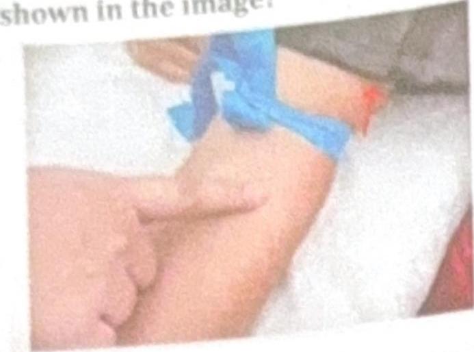
What is the name of the nerve block technique shown in the image?

Most commonly used approach of brachial plexus block?
Explanation: **Breast cancer** - Liver metastases from **breast cancer** frequently present as **hypoechoic lesions** on ultrasound, due to the tumor's cellular composition and vascularity. - This appearance helps differentiate them from other more commonly echogenic or mixed metastatic patterns. *RCC* - **Renal cell carcinoma (RCC)** metastases to the liver often appear **hyperechoic** or **mixed echogenicity** on ultrasound due to their rich vascularity. - This is a distinct characteristic, different from the predominantly hypoechoic nature seen with breast cancer metastases. *Colon cancer* - Liver metastases from **colon cancer** are typically **echogenic** or **mixed echogenicity** on ultrasound, sometimes with a hypoechoic rim ("target sign"). - Their presentation is generally not purely hypoechoic, making them distinguishable from breast cancer metastases. *Mucinous adenocarcinoma* - Liver metastases from **mucinous adenocarcinoma** can be quite variable, but they often appear **complex**, possibly with **cystic components** or mixed echogenicity, rather than uniformly hypoechoic. - The mucin content can create a distinct internal architecture on ultrasound that differs from solid hypoechoic lesions.
Explanation: ***USG*** - **Focused assessment with sonography for trauma (FAST) exam** is the investigation of choice in an **unstable patient** due to its rapid, non-invasive nature and ability to detect free fluid (blood) in the peritoneal, pericardial, and pleural spaces. - It can be performed at the **bedside** without moving the patient, making it ideal for hemodynamically unstable individuals with suspected intra-abdominal injury. *CT scan* - While a **CT scan** provides detailed anatomical information, it requires the patient to be stable enough for transport to a radiology suite and prolonged scanning time. - It is often difficult to obtain in **unstable patients** who may require continuous resuscitation and monitoring. *X-ray abdomen* - An **X-ray abdomen** has limited utility for detecting intra-abdominal injuries and primarily identifies issues like free air under the diaphragm (suggesting hollow organ perforation) or foreign bodies. - It is **not sensitive** for detecting free fluid (hemoperitoneum) or solid organ injuries, which are critical in trauma. *DPL* - **Diagnostic peritoneal lavage (DPL)** is an invasive procedure that involves inserting a catheter into the peritoneal cavity to detect blood or other fluid. - While sensitive, it is **invasive**, can complicate subsequent imaging, and has largely been replaced by the FAST exam due to the latter's non-invasive nature and comparable diagnostic accuracy for free fluid.
Explanation: ***SonoVue*** - **SonoVue** (also known as Lumason in the US) is the most commonly used **microbubble contrast agent** specifically designed for **ultrasonography (USG)**. - Contains **sulphur hexafluoride gas microbubbles** stabilized by a phospholipid shell. - These microbubbles enhance the echogenicity of blood, improving the visualization of blood flow and tissue perfusion in various organs, particularly in **liver imaging** and **cardiac echocardiography**. - Other USG contrast agents include Definity and Optison. *Urograffin* - **Urograffin** (diatrizoate) is an iodine-based contrast agent primarily used in **X-ray imaging**, particularly for **urography (imaging of the urinary tract)** and **angiography**. - It is not suitable for USG as it does not produce the necessary acoustic impedance difference for ultrasound enhancement. *Omnipaque* - **Omnipaque** (iohexol) is a non-ionic, low-osmolar **iodinated contrast agent** used for various **X-ray and CT procedures**. - Like other iodinated agents, its properties are optimized for X-ray attenuation, not for ultrasound enhancement. *Conray* - **Conray** (iothalamate) is an older generation, high-osmolar, **iodinated contrast agent** used for various **X-ray procedures** like angiography and CT scans. - Similar to Urograffin, it works by X-ray attenuation and is not effective for ultrasound contrast enhancement.
Explanation: ***INR > 2 in ophthalmic procedures*** - While a high INR (international normalized ratio) indicates increased bleeding risk, an INR > 2 is generally considered a **relative contraindication** for most regional anesthesia procedures, especially in ophthalmic cases where the risk of significant hemorrhage might be lower compared to deeper blocks. - The decision to proceed often depends on the specific procedure, patient's overall condition, and a careful risk-benefit analysis, rather than being an absolute bar. *Lack of resuscitation facilities* - The absence of proper **resuscitation equipment and trained personnel** is an absolute contraindication for regional anesthesia, as serious complications (e.g., local anesthetic systemic toxicity, respiratory depression) can occur, requiring immediate intervention. - Performing regional anesthesia in such a setting puts the patient at extreme risk of irreversible harm or death in the event of an adverse reaction. *Infection at injection site* - Performing regional anesthesia through an infected area carries a high risk of introducing bacteria into deeper tissues, including the **neuraxial space** or surrounding nerves, leading to serious complications like **abscess formation, meningitis, or osteomyelitis**. - This is an **absolute contraindication** to prevent the spread of infection. *Patient refusal* - **Informed consent** is a fundamental ethical and legal principle in medicine; a competent patient's refusal to undergo a procedure, including regional anesthesia, must always be respected. - Proceeding against a patient's wishes constitutes **assault and battery** and is an absolute contraindication for any medical intervention.
Explanation: ***Hydatid cyst*** - A **hydatid cyst** often presents on ultrasound as a **focal anechoic lesion** with characteristic **floating membranes** (water lily sign) due to detachment of the endocyst. - These cysts are caused by tapeworm larvae (*Echinococcus granulosus*) and can have daughter cysts within the main cyst. *Pyogenic abscess* - A **pyogenic abscess** typically appears as an **ill-defined, hypoechoic liver lesion** with internal debris, sometimes showing gas. - While it can be anechoic, the presence of **floating membranes** is not a characteristic feature. *Hemangioma* - A **hemangioma** is usually a **well-defined, hyperechoic lesion** on ultrasound, without any internal fluid or floating membranes. - Its appearance is distinct from cystic lesions and it often shows characteristic peripheral nodular enhancement on contrast-enhanced imaging. *Amoebic abscess* - An **amoebic abscess** typically presents as a **hypoechoic, often round or oval lesion** with fine internal echoes and poorly defined walls, usually without floating membranes. - It is frequently located in the right lobe of the liver and can be difficult to differentiate from a pyogenic abscess based on imaging alone.
Explanation: ***Surgical intervention*** - **Fat embolism syndrome (FES)** is a medical emergency primarily managed with **supportive care**, not surgery. - Surgical intervention is only indicated for the **initial injury**, such as stabilizing long bone fractures, which helps prevent fat emboli, but not for treating an already established FES [1]. *Oxygen therapy* - **Oxygen therapy** is a crucial component of FES management, as the syndrome often leads to **hypoxemia** due to lung involvement. - It helps maintain adequate **tissue oxygenation** and can be administered via nasal cannula, face mask, or mechanical ventilation in severe cases. *Heparin administration* - **Heparin administration** was historically used with the rationale of preventing thrombus formation and potentially breaking down fat globules. - However, its effectiveness is **unproven**, and it carries risks such as bleeding, so it is generally **not recommended** for FES. *Low Molecular Weight Dextran* - **Low Molecular Weight Dextran** has been investigated for its potential to improve blood flow, reduce fat globule aggregation, and expand plasma volume in FES. - While some studies showed promising results, it is **not a universally accepted standard treatment** due to conflicting evidence and potential side effects.
Explanation: ***Infraclavicular*** - The **infraclavicular approach** targets the brachial plexus at the level of the **cords**, which lie deep to the pectoralis muscles and medial to the coracoid process. - This block is particularly useful for surgeries involving the **distal upper limb**, as it blocks all divisions of the cords. *Supraclavicular* - The **supraclavicular approach** targets the brachial plexus at the level of the **trunks**, specifically where they emerge between the anterior and middle scalene muscles. - While it provides good anesthesia for the entire upper limb, it is proximal to the cord level. *Axillary* - The **axillary approach** targets the terminal nerves (e.g., median, ulnar, radial, musculocutaneous) after the brachial plexus has divided into individual nerves in the **axilla**. - This block is distal to the cords and is often used for surgeries of the forearm and hand. *All of the options* - This option is incorrect because only the **infraclavicular approach** specifically targets the cords of the brachial plexus. - The other approaches target either the trunks (supraclavicular) or the terminal nerves (axillary).
Explanation: ***Nasociliary nerve*** - A nasociliary nerve block specifically targets the **nasociliary nerve** itself. - This block is used to anesthetize the sensory innervation of structures supplied by the nasociliary nerve, such as parts of the **nasal cavity**, **eyeball**, and **skin of the nose**. *Greater palatine nerve* - The **greater palatine nerve** supplies sensation to the posterior hard palate and is targeted in a **greater palatine nerve block**. - This nerve is a branch of the **maxillary nerve** and is primarily involved in dental and palatal anesthesia. *Sphenopalatine nerve* - The **sphenopalatine nerve**, or pterygopalatine ganglion, contains sensory fibers for the nasal cavity, palate, and pharynx, and its block is distinct from a nasociliary block. - A **sphenopalatine ganglion block** is mainly used for conditions like cluster headaches and facial pain, not for direct eyeball sensation. *Anterior ethmoidal nerve* - The **anterior ethmoidal nerve** is a branch of the nasociliary nerve, but a nasociliary nerve block targets the main trunk, which includes all its branches. - While the anterior ethmoidal nerve supplies the anterior part of the nasal septum and lateral wall, it is a **component** of the nasociliary innervation rather than the sole target.
Explanation: ***Intra-arterial anesthesia*** - The image shows a **cannula inserted directly into an artery**, indicated by the blood reflux and the context of anesthesia, suggesting direct drug delivery into the arterial system. - This method is used for specific types of regional pain management or diagnostic procedures where direct arterial access is required for **localized drug distribution**. *Bier's block* - A Bier's block, or **intravenous regional anesthesia**, involves injecting local anesthetic into a **vein** in an extremity after it has been exsanguinated and isolated by a tourniquet. - The image clearly shows a **bright red blood flash**, characteristic of arterial cannulation, not venous. *Regional anesthesia* - This is a broad term referring to the **anesthesia of a specific region** of the body and encompasses various techniques. - While intra-arterial anesthesia is a type of regional anesthesia, "regional anesthesia" itself is too general to specifically describe the technique shown. *Axillary block* - An **axillary block** is a type of peripheral nerve block targeting the brachial plexus in the axilla to anesthetize the arm. - The image does not depict the axillary region or the characteristic needle placement for an axillary block; instead, it shows direct vascular access.
Explanation: ***Axillary*** - The **axillary approach** is the most frequently utilized technique for **brachial plexus block** due to its relatively superficial location, making it safer and easier to perform. - This approach is particularly effective for procedures involving the **forearm and hand**, as it reliably blocks the terminal branches of the brachial plexus. *Interscalene* - The **interscalene block** is primarily used for **shoulder surgery** as it provides excellent anesthesia to the shoulder, clavicle, and upper arm. - It carries a higher risk of complications like **phrenic nerve palsy** and **hoarseness** due to its proximity to vital structures. *Supraclavicular* - The **supraclavicular block** targets the **trunks** of the brachial plexus, making it suitable for procedures involving the **upper arm, forearm, and hand**. - This approach has a higher risk of **pneumothorax** due to its close proximity to the pleura. *Infraclavicular* - The **infraclavicular block** targets the **cords** of the brachial plexus, offering good anesthesia for surgeries of the **distal upper arm, forearm, and hand**. - It involves a deeper approach compared to axillary and is often guided by ultrasound to minimize risks, but is not as commonly used as the axillary for general procedures.
Get full access to all questions, explanations, and performance tracking.
Start For Free