Finkelstein test is used for diagnosis of?
A patient at the orthopedics OPD complains of troubled sleep at night due to numbness and tingling sensation involving his lateral 3 digits. His symptoms are relieved as he lays his arms hanging from the bed. Which of the following options correctly describes his condition and the test used to assess it?
Pointing index finger is seen in which nerve injury
Patient with shoulder dislocation has axillary nerve injury. Which movement will be most affected?
What is the characteristic upper limb deformity seen in Erb's palsy?
What is the condition commonly referred to as 'draughtsman's elbow'?
Ulnar nerve injury results in:
A 40-year-old man was repairing his wooden shed on Sunday morning. By afternoon, he felt that the hammer was becoming heavier and heavier. He felt pain in the lateral side of the elbow and also found that squeezing water out of sponge hurt his elbow. Which of the muscles are most likely involved-
Median nerve injury at the wrist causes -
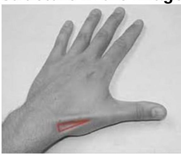
The image shows a highlighted region on the dorsal aspect of the hand (anatomical snuffbox). Which of the following anatomical structures form the boundaries or floor of this region?

Explanation: ***De quervain tenosynovitis*** - The **Finkelstein test** is a specific diagnostic maneuver for **De Quervain's tenosynovitis**, where a positive test elicits pain at the wrist. - This condition involves inflammation of the **extensor pollicis brevis** and **abductor pollicis longus tendons** within the first dorsal compartment of the wrist. *Tarsal tunnel syndrome* - This syndrome involves compression of the **posterior tibial nerve** in the ankle, not the wrist. - Diagnosis typically involves **Tinel's sign** over the tarsal tunnel and nerve conduction studies. *Carpal tunnel syndrome* - This condition involves compression of the **median nerve** at the wrist. - Diagnostic tests include **Phalen's maneuver** and **Tinel's sign** over the carpal tunnel, which differ from the Finkelstein test. *Thoracic outlet syndrome* - This involves compression of neurovascular structures in the **thoracic outlet**, typically affecting the neck and upper extremity but not the wrist specifically. - Diagnostic tests involve specific provocative maneuvers that assess for vascular or neurological compromise in the shoulder and arm.
Explanation: ***Carpal tunnel syndrome, Durkan's test*** - The symptoms of **numbness and tingling** in the **lateral 3 digits** (thumb, index, middle, and radial half of the ring finger) are classic for **carpal tunnel syndrome (CTS)**, caused by compression of the **median nerve**. Relief with hanging the arm is due to gravity reducing swelling and pressure. - **Durkan's test** (or **median nerve compression test**) is highly specific for CTS. It involves direct pressure over the carpal tunnel, reproducing symptoms within 30 seconds. *Guyon's canal syndrome, Froment's test* - **Guyon's canal syndrome** involves compression of the **ulnar nerve** at the wrist, primarily affecting the **little finger** and the **ulnar half of the ring finger**, not the lateral 3 digits. - **Froment's test** assesses **ulnar nerve palsy** by observing the strength of adductor pollicis during a pinch grip, which is unrelated to median nerve compression. *Carpal tunnel syndrome, Froment's test* - While **carpal tunnel syndrome** is correctly identified based on the symptoms, **Froment's test** is not used to assess it. - As mentioned, Froment's test evaluates **ulnar nerve function**, particularly the adductor pollicis muscle. *Guyon's canal syndrome, Durkan's test* - The symptoms described (lateral 3 digits) are inconsistent with **Guyon's canal syndrome**, which affects the ulnar nerve distribution. - Although **Durkan's test** is appropriate for carpal tunnel syndrome, the diagnosis for Guyon's canal syndrome is incorrect.
Explanation: ***Median*** - The **pointing index finger** sign occurs in **proximal median nerve injury** when attempting to make a fist, as the median nerve supplies **flexor digitorum superficialis (FDS)** and lateral half of **flexor digitorum profundus (FDP)**. - This creates the classic **"hand of benediction"** or **"pope's blessing"** sign where index and middle fingers remain extended due to loss of flexion capability. *Axillary* - Primarily innervates the **deltoid** and **teres minor muscles**, affecting **shoulder abduction** and external rotation. - Injury does not cause any **finger positioning abnormalities** or hand deformities. *Radial* - Injury causes **"wrist drop"** with inability to extend the wrist, thumb, and fingers at **MCP joints**. - Results in all fingers remaining **flexed due to unopposed flexor action**, not a pointing finger deformity. *Ulnar* - Injury causes **"claw hand"** deformity primarily affecting the **ring and little fingers** (4th and 5th digits). - Creates **hyperextension at MCP joints** and **flexion at PIP/DIP joints**, with the index finger typically less affected.
Explanation: ***Shoulder Abduction*** - The **axillary nerve** innervates the **deltoid muscle**, which is the primary muscle responsible for **shoulder abduction** beyond the initial 15 degrees. - Injury to this nerve would significantly impair the patient's ability to lift their arm away from their body. *Forward Flexion* - **Forward flexion** of the shoulder is primarily carried out by the **anterior deltoid**, **pectoralis major**, and **coracobrachialis muscles**. - While the anterior deltoid is affected, other muscles can still contribute to this movement, making it less severely impaired than abduction. *Internal Rotation* - **Internal rotation** is largely controlled by the **subscapularis**, **latissimus dorsi**, **teres major**, and **pectoralis major**. - These muscles are not innervated by the axillary nerve, so internal rotation would be largely preserved. *External Rotation* - **External rotation** is primarily performed by the **infraspinatus** and **teres minor muscles**. - These muscles are supplied by the **suprascapular nerve** and **axillary nerve** (for teres minor), respectively, but the deltoid's role is minimal, so overall external rotation would be less affected compared to abduction.
Explanation: ***Adduction and medial rotation of arm*** - Erb's palsy, resulting from injury to the **upper brachial plexus** (C5-C6 nerve roots), primarily affects the **deltoid**, **supraspinatus**, **infraspinatus**, and **biceps** muscles. - The unopposed action of unaffected muscles, such as the **pectoris major** and **latissimus dorsi**, leads to the characteristic **waiter's tip position**, involving **adduction** and **medial rotation** of the arm. *Adduction and lateral rotation of arm* - This position would imply weakness of the **pectoralis major** and **latissimus dorsi** and stronger activity of the **infraspinatus** and **teres minor**, which is contrary to the muscle deficits in Erb's palsy. - **Lateral rotation** of the arm is typically impaired in Erb's palsy due to weakness of the **infraspinatus** and **teres minor**. *Abduction and lateral rotation of arm* - **Abduction** is severely impacted in Erb's palsy due to paralysis of the **deltoid** and **supraspinatus**. - This position would suggest intact function of muscles that are explicitly weakened or paralyzed in Erb's palsy. *Abduction and medial rotation of arm* - While **medial rotation** can be a component of the deformity, **abduction** is a movement that is significantly impaired in Erb's palsy, making this option incorrect. - The inability to abduct the arm is a hallmark of the condition due to weakness of the **deltoid** and **supraspinatus**.
Explanation: ***Olecranon bursitis*** - This condition is colloquially known as **"draughtsman's elbow"**, **"student's elbow"**, or **"baker's elbow"** due to its association with prolonged leaning on the elbow. - It involves inflammation and swelling of the **olecranon bursa**, which is located at the posterior aspect of the elbow. *Lateral epicondylitis* - This condition is commonly known as **"tennis elbow"** and involves inflammation of the **extensor tendons** originating from the lateral epicondyle. - It typically presents with pain on the **lateral aspect of the elbow**, worsened by gripping and wrist extension. *Medial epicondylitis* - This condition is commonly known as **"golfer's elbow"** and involves inflammation of the **flexor-pronator tendons** originating from the medial epicondyle. - It causes pain on the **medial aspect of the elbow**, worsened by activities involving wrist flexion and pronation. *Medial epicondyle avulsion fracture* - This is a traumatic injury where a fragment of the **medial epicondyle** is pulled away from the humerus, often seen in overhead throwing athletes. - It usually involves acute pain, swelling, and sometimes **nerve dysfunction**, which differs significantly from the inflammatory process of olecranon bursitis.
Explanation: ***Clawing of fingers*** - An ulnar nerve injury, particularly at the elbow, often leads to **paralysis of the interossei muscles** and the **medial two lumbricals**. [1] - This results in **hyperextension at the metacarpophalangeal joints** and **flexion at the interphalangeal joints** of the 4th and 5th fingers (and sometimes 3rd), creating the characteristic claw hand deformity. [1] *Pointing index* - **Pointing index**, also known as the **sign of benediction** or **preacher's hand**, occurs with **high median nerve lesions** affecting the lateral lumbricals and flexor digitorum superficialis. - The patient is unable to flex the index and middle fingers, especially when attempting to make a fist. *Ape thumb deformity* - **Ape thumb deformity** is caused by a **median nerve injury**, specifically affecting the **thenar muscles** (abductor pollicis brevis, opponens pollicis, and superficial head of flexor pollicis brevis). - This paralysis leads to the thumb being pulled laterally and into the same plane as the other fingers, losing its ability to oppose. *Wrist drop* - **Wrist drop** is a classic sign of **radial nerve injury**, which paralyzes the **extensor muscles of the wrist and fingers**. - This prevents the patient from extending their wrist and metacarpophalangeal joints.
Explanation: ***Extensor carpi radialis longus and brevis*** - The symptoms described, such as **lateral elbow pain** and pain with actions like hammering and squeezing, are classic for **lateral epicondylitis**, also known as **tennis elbow**. - **Extensor carpi radialis longus** and **brevis** are the primary muscles that originate from the **lateral epicondyle**, and their tendons are commonly affected in this condition. *Triceps brachii and anconeus* - The **triceps brachii** is responsible for elbow extension; injury to this muscle or the anconeus would typically cause pain in the **posterior aspect of the elbow**. - Pain specifically localized to the **lateral elbow** with gripping and wrist extension activities is not characteristic of triceps or anconeus involvement. *Biceps brachii and supinator* - The **biceps brachii** is a primary supinator and elbow flexor, while the **supinator** muscle also aids in supination; involvement of these would typically cause pain in the **anterior elbow** or with supination against resistance. - These muscles are generally not associated with pain in the **lateral epicondyle** with wrist extension and gripping activities. *Flexor digitorum superficialis* - The **flexor digitorum superficialis** is involved in flexing the fingers and wrist and originates from the **medial epicondyle** of the humerus. - Injury to this muscle would cause pain on the **medial side of the elbow** (golfer's elbow), not the lateral side, and is typically exacerbated by repetitive wrist flexion.
Explanation: ***Loss of apposition of thumb*** - A **median nerve injury at the wrist** specifically affects the **motor branches to the thenar muscles**, including the **opponens pollicis, abductor pollicis brevis, and the superficial head of flexor pollicis brevis**. - This leads to an inability to **oppose the thumb** to the other fingers, significantly impairing fine motor skills and grasping. - Also causes sensory loss over the **lateral 3½ digits** (thumb, index, middle, and lateral half of ring finger). *Policeman's tip deformity* - This term is **not a standard clinical description** and may be confused with **waiter's tip hand** (Erb's palsy). - **Waiter's tip hand** results from injury to the **upper trunk of the brachial plexus (C5-C6)**, causing adduction and internal rotation of the shoulder with extension and pronation of the elbow. - This is a **completely different clinical picture** from median nerve injury at the wrist and involves proximal nerve injury, not peripheral nerve injury. *Saturday night palsy* - This condition is caused by **compression of the radial nerve** in the spiral groove of the humerus, often from prolonged pressure (e.g., falling asleep with an arm over a chair). - It results in **wrist drop** and impaired extension of the fingers and thumb, not specific thumb apposition issues. *Claw hand* - A claw hand deformity is typically caused by an injury to the **ulnar nerve** (affecting the medial two fingers more prominently) or a combined **median and ulnar nerve injury** (affecting all fingers). - It involves **hyperextension of the MCP joints** and **flexion of the IP joints** of the fingers, which is distinct from isolated thumb apposition loss seen in median nerve injury.
Explanation: ***All of the above anatomical structures.*** - The image highlights the **anatomical snuffbox**, a triangular depression on the radial dorsal aspect of the hand. Its boundaries are formed by the tendons of the **extensor pollicis longus muscle** (ulnar side), and the **abductor pollicis longus** and **extensor pollicis brevis muscles** (radial side). - The **styloid process of the radius** forms the floor of the anatomical snuffbox along with the scaphoid and trapezium bones. All the options listed are key anatomical features associated with this region. *Extensor pollicis longus muscle.* - This muscle forms the **ulnar (medial) border** of the anatomical snuffbox. - Its tendon can be palpated during **thumb extension** and contributes to the overall structure of the highlighted area. *Abductor pollicis longus muscle.* - This muscle, along with the extensor pollicis brevis, forms the **radial (lateral) border** of the anatomical snuffbox. - Its tendon is visible and palpable on the radial side of the highlighted region when the thumb is abducted. *Styloid process of the radius.* - This bony prominence is located at the **distal end of the radius** on the radial side of the wrist. - It forms part of the **proximal floor** of the anatomical snuffbox, contributing to its definition.
Get full access to all questions, explanations, and performance tracking.
Start For Free