The aortic hiatus is formed by the right and left crura of the diaphragm. Which of the following structures does NOT pass through the aortic hiatus?
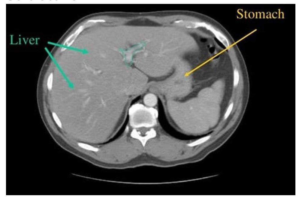
CT scan of abdomen showing a structure branching within the liver. Identify the structure.

When a lumbar puncture is performed to sample cerebrospinal fluid, which of the following external landmarks is the most reliable to determine the position of the L4 vertebral spine?
Which artery is the major supply of the medial surface of the cerebral hemisphere?
To visualize vascular sling causing tracheal or external airway compression, which of the following would you best prefer?
Which structure is not seen at the L3 level?
Blumensaat line is used to radiologically diagnose an anatomical condition called patella alta. It is
Which CT view is best for visualizing paranasal polyps?
Which type of study determines the odds ratio?
Which research method is most appropriate for studying the progression of a disease over time?
Explanation: ***Left gastric vein*** - The **left gastric vein** is part of the **portal venous system** and drains into the portal vein. - It **does NOT pass through the diaphragm** via the aortic hiatus or any other diaphragmatic opening. - It has **no anatomical relationship** with the aortic hiatus, making it the best answer to this question. *Thoracic duct* - The **thoracic duct** is the largest lymphatic vessel in the body and **passes through the aortic hiatus** along with the aorta. - It ascends through the aortic hiatus at the **T12 vertebral level** to eventually drain into the left subclavian vein. - It lies posterior to the aorta as it traverses the hiatus. *Left vagus nerve* - The **left vagus nerve** does NOT pass through the aortic hiatus, but it **does pass through the esophageal hiatus** at the T10 level. - It contributes to the **anterior vagal trunk** as it enters the abdomen with the esophagus. - While this structure doesn't pass through the aortic hiatus, it does traverse the diaphragm through a different opening, making it a less definitive answer than the left gastric vein. *Azygos vein* - The **azygos vein** typically **passes through the aortic hiatus** alongside the aorta and thoracic duct. - It may occasionally pass through a separate opening in the right crus of the diaphragm. - It collects deoxygenated blood from the posterior walls of the thorax and abdomen before draining into the superior vena cava.
Explanation: ***Portal vein*** - The image shows a **branching vessel within the liver parenchyma**. The **portal vein** enters the liver at the porta hepatis and branches extensively to supply the liver with nutrient-rich, deoxygenated blood from the gastrointestinal tract. - On a CT scan, the portal vein and its branches appear as prominent, contrast-filled structures centrally located within the liver, consistent with the identified structure. *Superior Vena Cava* - The **superior vena cava** is located in the **chest**, superior to the diaphragm, and drains blood from the upper body into the right atrium; it does not branch within the liver. - This vessel would not be visible in an abdominal CT slice at this level and does not show intrahepatic branching. *Inferior Vena Cava* - The **inferior vena cava (IVC)** is a large vessel located **posterior to the liver**, collecting deoxygenated blood from the lower body and liver (via hepatic veins) before emptying into the right atrium. - While it is in the abdomen, it does not branch within the liver parenchyma in the same manner as the portal vein; rather, **hepatic veins** drain into it from the liver. *Splenic Vein* - The **splenic vein** runs along the **posterior aspect of the pancreas** and eventually joins with the superior mesenteric vein to form the portal vein outside the liver. - It does not enter or branch within the liver itself; its location is too far posterior and outside the liver to match the structure indicated.
Explanation: ***The iliac crests*** - A line drawn between the **highest points of the iliac crests** on both sides typically intersects the L4 vertebral body or the L4-L5 intervertebral space. - This anatomical landmark provides a **safe entry point** for lumbar puncture, avoiding the spinal cord which usually ends at L1-L2. *The lowest pair of ribs bilaterally* - The lowest pair of ribs (12th ribs) corresponds to the **twelfth thoracic vertebra (T12)**, which is much higher than the desired lumbar puncture site. - Using this landmark would place the needle at a level where the **spinal cord is still present**, posing a significant risk of injury. *The inferior angles of the scapulae* - The inferior angle of the scapula typically corresponds to the **seventh thoracic vertebra (T7)**. - This landmark is also too superior for a safe lumbar puncture and does not accurately localize the lumbar spine. *The posterior superior iliac spines* - The posterior superior iliac spines (PSIS) are located at the level of the **S2 vertebra**, which is too far inferior for a standard lumbar puncture at L4-L5. - While they are important pelvic landmarks, they are not used for determining the L4 vertebral spine in this context.
Explanation: ***Anterior cerebral artery*** - The **anterior cerebral artery (ACA)** is a primary branch of the internal carotid artery and is responsible for supplying blood to the **medial surface** of the frontal and parietal lobes of the cerebral hemispheres [1]. - It also supplies the **corpus callosum**, the superior aspect of the frontal and parietal lobes, and parts of the basal ganglia [1]. *Posterior cerebral artery* - The **posterior cerebral artery (PCA)** primarily supplies the **occipital lobe** and the inferior part of the **temporal lobe** [1]. - It also provides blood to parts of the midbrain and the **thalamus** [1]. *Middle cerebral artery* - The **middle cerebral artery (MCA)** is the largest cerebral artery and supplies most of the **lateral surface** of the cerebral hemispheres [1]. - It is crucial for the blood supply to the **motor and sensory cortices** for the face and upper limb, as well as language areas (Broca's and Wernicke's). *Posterior inferior cerebellar artery* - The **posterior inferior cerebellar artery (PICA)** is a branch of the **vertebral artery** and exclusively supplies the **cerebellum** and the lateral medulla. - It is not involved in the blood supply to the cerebral hemispheres.
Explanation: ***Correct: CT (CT Angiography)*** - **CT angiography is the gold standard** for diagnosing vascular rings and slings causing airway compression - Provides **excellent spatial resolution** with multiplanar and 3D reconstruction capabilities to clearly demonstrate the anatomical relationship between anomalous vessels and the trachea/bronchi - **Fast acquisition time** minimizes motion artifacts, particularly important in pediatric patients who are the typical population affected by vascular slings - Superior for **surgical planning** due to detailed visualization of vascular anatomy and the exact site and degree of airway compression - More readily available and cost-effective compared to MRI for this specific indication *Incorrect: MRI* - While MRI provides excellent soft tissue contrast and avoids ionizing radiation, it has **longer acquisition times** leading to increased risk of motion artifacts, especially in children - Lower spatial resolution compared to CT for vascular structures - May be used as a **complementary modality** when radiation avoidance is critical or for follow-up imaging, but not the first-line preferred modality *Incorrect: Catheter angiography of aorta and pulmonary artery* - **Invasive procedure** with associated risks including vascular injury, bleeding, and contrast reactions - Provides excellent vascular detail but **no information about airway compression** or surrounding soft tissue structures - Reserved for cases requiring **intervention** (embolization, stent placement) or when non-invasive imaging is inconclusive - Has been largely replaced by non-invasive CTA for diagnostic purposes *Incorrect: PET-CT* - Primarily assesses **metabolic activity**, used for oncology staging, infection, and inflammatory conditions - **Not indicated** for structural vascular anomalies or their anatomical relationships to airways - Does not provide the necessary vascular or airway detail for evaluating vascular slings
Explanation: ***Coeliac trunk*** - The **coeliac trunk** typically arises from the abdominal **aorta** at the level of **T12-L1**, which is significantly higher than L3. - It then immediately branches into the **left gastric**, **splenic**, and **common hepatic arteries** to supply foregut structures. *Iliac vessels* - The **common iliac arteries** and veins typically bifurcate from the **aorta** and **IVC** around the L4-L5 level. - Their presence, or the start of their formation, can be observed near or just above **L3**, depending on individual anatomical variation and how "at the L3 level" is interpreted (e.g., within the L3 vertebral body's span). *Aorta* - The **abdominal aorta** descends along the posterior abdominal wall and is a prominent structure at the **L3 level**. - It typically bifurcates into the common iliac arteries at the level of **L4**, meaning it is still a large, undivided vessel at L3. *IVC* - The **inferior vena cava (IVC)** ascends through the abdomen and is a significant vascular structure at the **L3 level**. - It is formed by the union of the common iliac veins at the level of **L5** and continues superiorly.
Explanation: ***Drawn through the roof of the intercondylar notch of femur*** - The **Blumensaat line** is a radiologic landmark defined by the **roof of the intercondylar notch** of the femur on a lateral knee X-ray. - It is used in the diagnosis of **patella alta** or **patella baja** by assessing the position of the patella relative to this line; the inferior pole of the patella should ideally be at or slightly below the Blumensaat line in a flexed knee. *The line joining the greater trochanter and the centre of the patella* - This description does not correspond to the **Blumensaat line** or any standard anatomical measurement for patellar position. - The greater trochanter is part of the proximal femur, far removed from the knee joint for this specific radiological evaluation. *Drawn through the superior pole of patella.* - While lines can be drawn in relation to the patella, the **Blumensaat line** is specifically defined by the **femoral intercondylar notch**, not the superior pole of the patella. - Other indices like the **Insall-Salvati ratio** use the patellar pole, but in relation to the patellar tendon length, not as the Blumensaat line itself. *The line joining the ASIS with the centre of patella* - This line is not the **Blumensaat line** and does not have a standard application in diagnosing **patella alta** or baja. - The **anterior superior iliac spine (ASIS)** is part of the pelvis and would not be used in a direct measurement of patellar height on a knee X-ray.
Explanation: ***Coronal*** - The **coronal view** provides the best visualization of the **ostia of the paranasal sinuses**, which are crucial for assessing the extent and obstruction caused by polyps. - This orientation effectively demonstrates whether polyps are **protruding into the nasal cavity** or obstructing the drainage pathways. *Axial view* - The axial view is useful for evaluating **posterior structures** and **bony erosion** but is less optimal for assessing the vertical extent of polyps or ostial obstruction. - It can show the **anteroposterior dimensions** of polyps but does not offer the same clarity for sinus outflow tracts as the coronal view. *Sagittal view* - The sagittal view is good for showing the **craniocaudal extent** of lesions and differentiating between the nasal cavity and sphenoid sinus, but it is not ideal for comprehensive paranasal sinus polyp evaluation. - It can help in localizing some polyps but does not provide a clear overview of **sinus ostia** or lateral extension. *3D view* - A 3D reconstruction can be helpful for a general overview and surgical planning but does not offer the fine detail and specific orientation needed for primary polyp detection and ostial assessment as effectively as direct 2D views. - It is a derived image rather than a primary acquisition plane and might obscure smaller polyps or subtle anatomical relationships.
Explanation: ***Case control*** - **Case-control studies** compare individuals with a disease (cases) to individuals without the disease (controls) and look back in time to identify previous exposures. - The **odds ratio** is the primary measure of association used in case-control studies, quantifying the odds of exposure among cases versus controls. *Cohort* - **Cohort studies** follow groups of individuals over time, some exposed to a risk factor and some not, to determine the incidence of a disease. - They typically determine **relative risk**, which is the ratio of incidence rates in exposed versus unexposed groups. *Cross sectional* - **Cross-sectional studies** assess the prevalence of disease and exposure at a single point in time. - They primarily measure **prevalence** and can be used to calculate a **prevalence odds ratio**, but they do not establish temporality between exposure and outcome. *RCT* - **Randomized controlled trials (RCTs)** are interventional studies where participants are randomly assigned to an intervention or control group to determine the effectiveness of a treatment or exposure. - The main measure of effect in RCTs is often the **relative risk reduction**, **absolute risk reduction**, or **number needed to treat**, rather than the odds ratio for observational exposure.
Explanation: ***Cohort Study*** - A **cohort study** observes a group of individuals over a period of time, allowing researchers to track the **natural progression of a disease** from exposure through onset and various stages. - This design is ideal for investigating the **incidence** of disease and identifying risk factors over time. *Cross sectional study* - A **cross-sectional study** assesses exposure and outcome at a **single point in time**, providing a snapshot. - It cannot establish temporality or observe disease progression, as it does not follow individuals over time. *Randomized Control Trials* - **Randomized controlled trials (RCTs)** are primarily designed to evaluate the **effectiveness of interventions** or treatments by comparing outcomes between an experimental group and a control group. - While they follow participants over time, their main goal is not to study the natural progression of an untreated disease. *Interventional Studies* - **Interventional studies** involve manipulating an exposure or treatment to observe its effect, often to test a hypothesis about a causal relationship. - While they track outcomes over time, their focus is on the impact of the intervention rather than the natural history or progression of a disease without intervention.
Get full access to all questions, explanations, and performance tracking.
Start For Free