Most abundant collagen in the body is
Which of the following statements is NOT true regarding red muscle fibers?
What is the order of bands in a sarcomere from the Z-disc toward the center?
Type I collagen is present in all EXCEPT:
Dense irregular connective tissue is found in:
Which of the following is NOT a glial cell?
All the following features are seen in neurons from dorsal root ganglia, EXCEPT:
Which type of collagen is most abundant in hyaline cartilage?
Type I collagen is present in all EXCEPT:
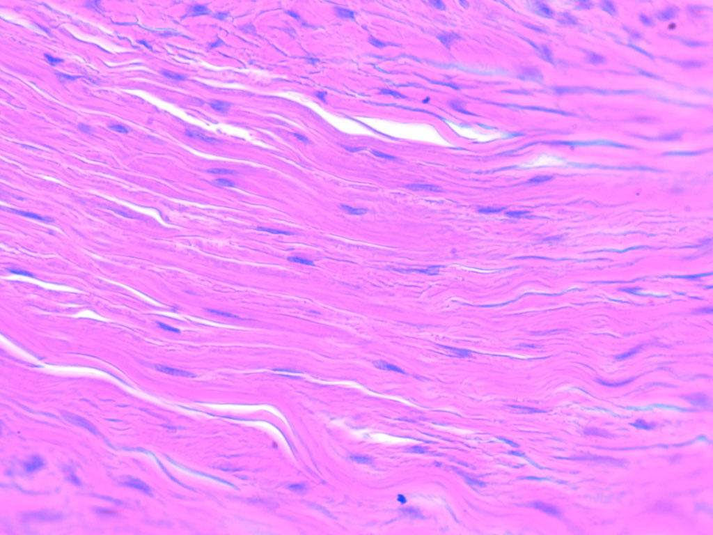
Identify the type of connective tissue present in the image.

Explanation: ***Type I*** - **Type I collagen** is the most abundant type in the human body, constituting about 90% of the body's total collagen. - It is primarily found in **skin, tendons, ligaments, bone, dentin, and intervertebral discs**, providing mechanical strength and structural integrity. *Type II* - **Type II collagen** is the main collagen found in **cartilage**, especially hyaline and elastic cartilage. - It provides resistance to pressure and is crucial for the structure of the **intervertebral disc nucleus pulposus** and the **vitreous humor of the eye**. *Type V* - **Type V collagen** is a minor fibrillar collagen that associates with **type I collagen** to regulate fibril diameter and organization. - It is found in **cornea, bone, and interstitial matrices**, playing a role in tissue development and integrity. *Type VI* - **Type VI collagen** is a microfibrillar collagen that forms bead-like microfibrils and is found in most **interstitial tissues**. - It plays a significant role in anchoring other extracellular matrix components and is particularly abundant in the **basement membranes** of blood vessels and muscles.
Explanation: ***Increased muscle fiber length*** - The length of muscle fibers is generally determined by the muscle's anatomical structure and function, not by whether they are red or white fibers. - While red muscle fibers (slow-twitch) are optimized for **endurance** and **sustained contractions**, this optimization does not involve an inherent increase in the length of individual muscle fibers. *Decreased glycolytic enzymes* - Red muscle fibers, also known as slow-oxidative fibers, primarily rely on **aerobic metabolism** for ATP production. - They have a lower content of glycolytic enzymes compared to white muscle fibers, which are specialized for **anaerobic glycolysis**. *Increased blood flow* - Red muscle fibers have a rich capillary supply, leading to **increased blood flow**, which is essential for delivering oxygen and nutrients for sustained aerobic activity. - This extensive vascularization contributes to their characteristic red appearance and their ability to resist fatigue. *Used for aerobic activity* - Red muscle fibers are well-suited for prolonged, low-intensity activities due to their high concentration of **mitochondria**, myoglobin, and oxidative enzymes. - They are primarily responsible for maintaining posture and performing **endurance activities** such as long-distance running.
Explanation: ***Z-A-H-M*** - This sequence accurately represents the arrangement of bands within a **sarcomere** when moving from the **Z-disc** towards the central **M-line**. - The **Z-disc** anchors **actin (thin) filaments**, which extend into the **A-band**, partially overlapping with myosin (thick) filaments. The **H-zone** is within the A-band, and the **M-line** bisects the H-zone. *Z-M-A-H* - This order incorrectly places the **M-line** immediately after the **Z-disc** and before the A and H bands. - The **M-line** is located at the very center of the sarcomere, a significant distance from the Z-disc. *Z-H-A-M* - This sequence incorrectly places the **H-zone** before the entire **A-band**. - The **H-zone** is a region *within* the **A-band**, specifically where only myosin (thick) filaments are present without actin (thin) overlap. *Z-H-M-A* - This order incorrectly places the **H-zone** and **M-line** before the **A-band**. - The **A-band** encompasses the entire length of the myosin (thick) filaments and includes the **H-zone** and **M-line** centrally.
Explanation: Cartilage - **Type II collagen** is the predominant collagen found in hyaline and elastic cartilage (the typical forms of cartilage), providing their characteristic tensile strength and resilience [2]. - Type I collagen is NOT the primary collagen in cartilage, making this the correct answer. - Note: Fibrocartilage is a specialized form that does contain Type I collagen, but standard cartilage refers to hyaline and elastic types. *Ligament* - **Type I collagen** is the primary structural component of ligaments, providing high tensile strength to connect bones and stabilize joints. - Its presence allows ligaments to withstand significant pulling forces without stretching excessively. *Aponeurosis* - **Type I collagen** is abundant in aponeuroses, which are flat sheet-like tendons that connect muscles to bones or other muscles. - This type of collagen provides the necessary tensile strength for these broad connective tissues. *Bone* - **Type I collagen** is the most abundant collagen in bone matrix, accounting for approximately 90% of its organic content [1]. - It forms a robust scaffold that gives bone its flexibility and tensile strength, working in conjunction with mineralized components like hydroxyapatite [1].
Explanation: ***Dermis*** - The **dermis** is primarily composed of **dense irregular connective tissue**, which provides structural strength and elasticity to the skin. [1] - This tissue consists of a rich network of **collagen fibers** arranged in various directions, along with elastic fibers, offering resistance to stress from multiple directions. [1] *Ligament* - Ligaments are examples of **dense regular connective tissue**, not irregular. - Their collagen fibers are densely packed and arranged in a **parallel fashion** to resist unidirectional stress. *Lamina propria* - The lamina propria is a layer of **loose connective tissue** underlying the epithelium of many organs. - It contains a higher proportion of **ground substance** and fewer, more loosely arranged fibers compared to dense connective tissue. *Tendon* - Tendons are also examples of **dense regular connective tissue**, connecting muscle to bone. - They are characterized by **parallel arrays of collagen fibers** adapted to withstand strong tensile forces in a single direction.
Explanation: ***Neurons*** - **Neurons** are the primary cells responsible for transmitting electrical and chemical signals throughout the nervous system [3]. - They are distinctly different from glial cells, which primarily provide **support and protection** to neurons [1]. *Oligodendrocytes* - **Oligodendrocytes** are a type of glial cell found in the **central nervous system** (CNS) [1]. - Their main function is to produce **myelin sheaths** that insulate axons, enhancing the speed of nerve impulse transmission [2]. *Microglia* - **Microglia** are the resident **immune cells** of the central nervous system, acting as its primary form of **active immune defense** [1]. - They scavenge for plaques, damaged neurons, and infectious agents, playing a critical role in neuroinflammation and tissue repair [2]. *Astrocytes* - **Astrocytes** are star-shaped glial cells found in the **brain and spinal cord** [1]. - They perform numerous functions, including providing **nutritional support**, maintaining the **blood-brain barrier**, and regulating the **extracellular environment** by controlling ion concentrations.
Explanation: ***They are multipolar*** - Dorsal root ganglia (DRG) neurons are typically **pseudounipolar**, meaning they have a single process that branches into two (peripheral and central) rather than multiple dendrites and an axon [1]. - **Multipolar neurons** are characteristic of motor neurons and interneurons in the central nervous system, not DRG sensory neurons [1]. *They contain lipofuscin granules* - **Lipofuscin granules** are common in long-lived, post-mitotic cells like neurons and are considered "wear and tear" pigments, accumulating with age. - Their presence in DRG neurons is a normal finding and reflects the neuron's metabolic activity over time. *They have eccentrically located nuclei* - While not universally present in all DRG neurons, an **eccentrically located nucleus** is a common histological feature of certain types of DRG neurons, particularly larger ones. - This feature helps distinguish them from other neuron types and can be accentuated by the large amount of cytoplasm in these cells. *They are derived from neural crest cells* - All sensory neurons of the DRG, along with other components like Schwann cells and sympathetic ganglia, originate from **neural crest cells**. - This developmental origin is a fundamental characteristic of DRG neurons, distinguishing them from CNS neurons derived from the neural tube.
Explanation: ***Type II*** - **Type II collagen** is the predominant type found in **hyaline cartilage**, providing tensile strength and elasticity [1]. - It is crucial for the **structural integrity** and functionality of cartilage in articular surfaces [1]. *Type I* - Predominantly found in **bone**, tendons, and skin, contributing to tensile strength but not a major component of hyaline cartilage [2]. - It forms the structure of **fibrocartilage**, such as in the **intervertebral discs**. *Type IV* - Mainly located in **basement membranes** and plays a role in filtration and structural support of epithelial cells, not in hyaline cartilage. - It is critical in the formation of structures like **glomeruli** in kidneys, differing from cartilage's needs. *Type III* - Found in **reticular fibers** and supporting tissues throughout the body, important for organ structure but not prominent in hyaline cartilage. - Often associated with **vascular structures** and is not involved in the composition of cartilage.
Explanation: ***Cartilage*** - **Type II collagen** is the predominant collagen found in hyaline and elastic cartilage, providing its characteristic tensile strength and resilience. - While some specialized cartilages like fibrocartilage contain Type I collagen, it is not the primary or universal type of collagen in cartilage overall. *Bone* - **Type I collagen** is the most abundant protein in bone [1], forming the organic matrix upon which mineral crystals (hydroxyapatite) are deposited [2]. - This collagen provides bone with its essential **tensile strength** and flexibility. *Ligament* - **Type I collagen** is the primary structural component of ligaments, contributing significantly to their ability to withstand high tensile forces. - Ligaments connect bones to bones, and the dense, ordered arrangement of Type I collagen fibers provides their crucial **mechanical stability**. *Aponeurosis* - **Type I collagen** is the main collagen type found in aponeuroses, which are broad, flat sheets of fibrous connective tissue that serve as attachments for muscles. - Similar to tendons, the strong, parallel arrangement of Type I collagen fibers allows aponeuroses to transmit **force efficiently**.
Explanation: ***Dense irregular*** - The image shows **densely packed collagen fibers** arranged in an **irregular, haphazard fashion**, characteristic of dense irregular connective tissue. - This type of tissue provides **strength** and resistance to stress from multiple directions, found in the **dermis of the skin** and organ capsules. *Loose and irregular* - **Loose connective tissue** would show more ground substance and fewer, loosely arranged fibers, whereas this image displays high fiber density. - While it is "irregular" in fiber arrangement, the density of fibers rules out the "loose" classification. *Specialized* - **Specialized connective tissues** include cartilage, bone, blood, and adipose tissue, which have distinct cellular and extracellular matrix components not seen here. - This tissue lacks the specific cellular and matrix characteristics that would classify it as specialized (e.g., chondrocytes in lacunae, osteocytes, blood cells). *Dense regular* - **Dense regular connective tissue** features collagen fibers arranged in parallel bundles, providing strength in one direction. - Examples include **tendons and ligaments**, which are structurally organized in an orderly, aligned manner, unlike the displayed irregular arrangement.
Get full access to all questions, explanations, and performance tracking.
Start For Free