Most common fractured facial bone
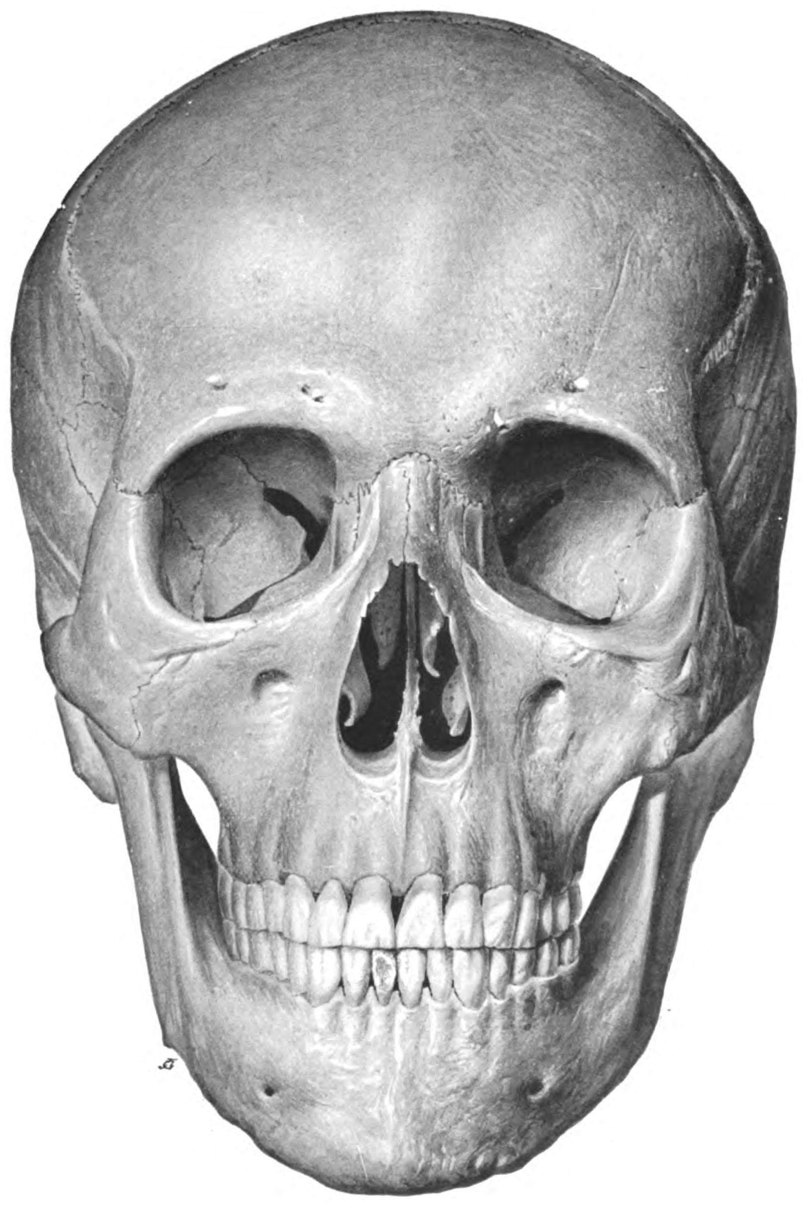
True statement about the skull shown below:

Le Fort II facial fracture implies:
Foramen spinosum transmits which of the following structures?
The most common bone involved in facial fractures is:
All of the following structures lie outside the cavernous sinus except:
A 43-year-old man presents to the emergency department after falling down a flight of stairs and landing on his head. He did not lose consciousness. He complains of severe headache, marked decreased acuity in hearing in the left ear, and a "runny nose" since the fall. On physical examination, he is found to have a left-sided Battle's sign (an ecchymosis in the area of the left mastoid process) and hemotympanum. He has a constant dripping of a clear, watery fluid through his nose. Findings on his neurologic examination, other than the hearing loss, are completely normal. X-ray studies will reveal which of the following?
Cranial nerve VIII passes through which of the following?
Paresthesia is seen with which of the following types of fractures:
A 25-year-old male presents with a head injury following a motorcycle accident, and a CT scan shows a fracture of the skull. Which bone is most commonly fractured in such injuries?
Explanation: ***Nasal bone*** - The **nasal bone** is the **most commonly fractured facial bone** due to its prominent and anterior position on the face. - Its relatively thin and delicate structure makes it highly susceptible to direct trauma, especially during sports injuries, falls, or assaults. *Nasoethmoid bone* - Fractures of the **nasoethmoid complex** are serious but less frequent than isolated nasal bone fractures, often resulting from high-impact trauma. - These fractures typically involve the **nasal bones**, **ethmoid sinuses**, and sometimes the medial orbital walls, leading to complex midfacial injuries. *Zygomatic bone* - The **zygomatic bone (cheekbone)** is the second most commonly fractured facial bone, but not the first. - Zygomatic fractures often occur from direct blows to the cheek but require more force than nasal bone fractures due to its sturdier structure. *Mandible* - The **mandible (jawbone)** is a robust bone, and while mandibular fractures are common facial injuries, they are secondary to nasal bone fractures in terms of frequency. - Mandibular fractures often result from significant force, such as motor vehicle accidents or direct blows to the jaw.
Explanation: ***Prominent supraorbital ridges, prominent glabella, square chin, acute fronto-nasal angle - Male*** - The image displays features typical of a **male skull**, including a pronounced **supraorbital ridge** (brow ridge), a prominent **glabella** (area between the eyebrows), a more **square and robust chin**, and an **acute fronto-nasal angle**. - These characteristics reflect the usually heavier and denser bone structure in males, adapted for stronger muscle attachments. *Round shaped orbit, prominent frontal eminences - Female* - **Female skulls generally have rounder orbits** and more prominent frontal eminences, giving the forehead a fuller, more rounded appearance. - The skull in the image does not exhibit these characteristics; the orbits are squarer, and the frontal eminences are not particularly prominent. *Prominent supraorbital ridges, prominent glabella, square chin, acute fronto-nasal angle - Female* - This statement incorrectly attributes features typically found in **male skulls** to a female skull. - Female skulls are generally smoother and less rugged, lacking the prominently developed ridges and angles seen in the image. *Round shaped orbit, prominent frontal eminences - Male* - This statement incorrectly attributes features typical of **female skulls** to a male skull. - Male skulls usually have squarer orbits and less prominent frontal eminences, with a more sloping forehead compared to females.
Explanation: ***Fracture running through zygomatic process of the maxilla, floor of orbit, and root of nose bilaterally*** - A **Le Fort II fracture**, also known as a **pyramidal fracture**, involves the separation of the midface from the cranium. - The fracture line typically extends bilaterally from the **nasal bones** through the **lacrimal bones**, **orbital floors**, and **zygomaticomaxillary sutures**, involving the **zygomatic process of the maxilla**. *Fracture running through alveolar ridge* - This description is characteristic of a **Le Fort I fracture**, which is also known as a **transverse maxillary fracture**. - A **Le Fort I fracture** involves separation of the palate and alveolar processes from the rest of the maxilla at the level of the nasal floor. *Fracture running through midline of the palate and zygomatico-maxillary suture* - While Le Fort fractures can involve the **zygomaticomaxillary suture**, a fracture specifically through the **midline of the palate** is more indicative of a **palatal fracture** or can be a component of a **Le Fort I fracture** if it extends transversely. - The unique combination described (midline palate and zygomatico-maxillary suture) does not perfectly fit the established Le Fort classifications on its own. *Bilateral fracture involving multiple facial bones with midface mobility* - While there is **midface mobility** in most Le Fort fractures, this description is too generic and could apply to **Le Fort II** or **Le Fort III fractures**. - It does not specify the precise anatomical path of the fracture, which is crucial for distinguishing between the different Le Fort types.
Explanation: ***Middle meningeal artery*** - The **foramen spinosum**, a small opening in the greater wing of the sphenoid bone, is primarily known for transmitting the **middle meningeal artery**. - This artery supplies blood to the **dura mater** and calvaria, making its passage through the foramen spinosum crucial for intracranial arterial supply. *Lateral petrosal nerve* - The **lesser petrosal nerve**, not the lateral petrosal nerve, passes through the **foramen ovale** or occasionally a small fissure near it, not the foramen spinosum. - The lesser petrosal nerve is involved in **parasympathetic innervation** to the parotid gland. *Mandibular nerve* - The **mandibular nerve** (V3), a branch of the trigeminal nerve, exits the skull through the **foramen ovale**, located anterior and lateral to the foramen spinosum. - It provides both **sensory and motor innervation** to structures in the head and neck. *Maxillary nerve* - The **maxillary nerve** (V2), another branch of the trigeminal nerve, exits the skull through the **foramen rotundum**, which is anterior to the foramen ovale. - It is primarily responsible for **sensory innervation** to the midface region.
Explanation: ***Mandible*** - The **mandible** is the most frequently fractured facial bone due to its prominent and exposed position, making it highly susceptible to trauma. - Common mechanisms of injury include **motor vehicle accidents**, falls, and assaults. *Zygomatic* - Although the **zygoma** (cheekbone) is commonly fractured, it ranks second or third after the mandible and nasal bones. - Fractures of the zygoma often involve displacement of the **zygomaticomaxillary complex (ZMC)**. *Nasal bones* - **Nasal bone fractures** are very common due to their delicate structure and exposed location on the face. - However, in terms of overall facial fractures, the **mandible** is more frequently involved. *Maxilla* - **Maxillary fractures**, often classified by Le Fort patterns, are less common than mandibular or nasal bone fractures. - These fractures typically result from **high-impact trauma** and can involve significant midfacial disruption.
Explanation: ***Internal carotid artery*** - The **internal carotid artery** passes directly through the **venous cavity** of the **cavernous sinus**, surrounded by venous blood. - This anatomical relationship is clinically significant, as trauma to the ICA within the sinus can lead to a **carotid-cavernous fistula**. - The ICA and the **abducent nerve (CN VI)** are the only structures that lie freely within the venous blood of the cavernous sinus. *Sphenoidal air sinus* - The **sphenoidal air sinus** is an air-filled cavity located inferior and anterior to the **cavernous sinus**, separated by a thin bony wall. - It is a distinct anatomical structure completely outside the cavernous sinus. *Maxillary nerve* - The **maxillary nerve (V2)** is located in the **lateral wall** of the cavernous sinus, embedded within the dura mater. - Unlike the ICA, it does **not** lie within the venous cavity itself, but rather within the thickness of the lateral wall. - It exits the skull through the **foramen rotundum** to enter the pterygopalatine fossa. - For the purposes of this question, structures in the lateral wall are considered separate from those within the venous space. *Foramen lacerum* - The **foramen lacerum** is an opening in the base of the skull, inferomedial to the **cavernous sinus**. - It is a bony aperture, not a structure within the cavernous sinus itself. - No major structures fully traverse the foramen lacerum as a completed entity in adults; instead, it is largely filled with fibrocartilage.
Explanation: ***A temporal bone fracture with CSF rhinorrhea*** - The combination of **Battle's sign**, **hemotympanum**, unilateral hearing loss, and clear nasal discharge after head trauma strongly indicates a **temporal bone fracture**. - **CSF rhinorrhea** refers to cerebrospinal fluid leaking from the nose due to a skull base fracture involving the temporal bone, typically affecting the petrous part. - The CSF can reach the nasal cavity via the **eustachian tube** or through fracture lines extending to the middle ear and mastoid air cells. *Occipital bone fracture* - While occipital fractures are possible with head trauma, they do not directly explain the specific findings of **hemotympanum** or unilateral hearing loss. - An occipital fracture would typically cause symptoms related to damage to the **brainstem** or **cerebellum**, depending on the extent. *A skull-base fracture with a mucocele* - A **mucocele** is a cyst filled with mucus, usually resulting from obstruction of a sinus ostium, and is not an acute traumatic finding. - While a skull-base fracture is present, the presence of a mucocele does not fit the acute injury presentation. *A fracture of the cribriform plate with a CSF leak into the paranasal sinuses* - A **cribriform plate fracture** would result in CSF rhinorrhea, but it typically causes CSF to leak directly from the anterior cranial fossa into the nasal cavity. - It would not explain the **hemotympanum**, Battle's sign, or unilateral hearing loss, which are characteristic of **temporal bone injury**.
Explanation: ***Internal acoustic meatus*** - The **internal acoustic meatus** is the bony canal that transmits the **vestibulocochlear nerve (CN VIII)**, along with the **facial nerve (CN VII)** and the **labyrinthine artery**, from the posterior cranial fossa to the inner ear [1]. - This passageway is crucial for the functions of hearing and balance mediated by CN VIII [1]. *Foramen ovale* - The **foramen ovale** transmits themandibular nerve **(CN V3)**, which is a branch of the trigeminal nerve, and is involved in mastication and sensory innervation of the lower face. - It also allows passage of the **accessory meningeal artery** and the **emissary veins**. *Foramen rotundum* - The **foramen rotundum** is a passage for the **maxillary nerve (CN V2)**, another branch of the trigeminal nerve, providing sensory innervation to the midface. - It primarily connects the middle cranial fossa with the pterygopalatine fossa. *Stylomastoid foramen* - The **stylomastoid foramen** is the exit point for the **facial nerve (CN VII)** from the skull, after it has passed through the internal acoustic meatus and the facial canal. - It is located between the styloid and mastoid processes of the temporal bone, allowing the facial nerve to emerge and innervate the muscles of facial expression.
Explanation: ***Zygomatico maxillary*** - Fractures involving the **zygomatico maxillary complex** (ZMC) can damage the **infraorbital nerve**, which passes through the infraorbital canal within the maxilla part of the ZMC. - Damage to the infraorbital nerve results in **paresthesia** (numbness or tingling) in the distribution of this nerve, affecting the cheek, upper lip, and anterior maxillary teeth on the affected side. *Coronoid process* - Fractures of the **coronoid process** are generally stable and typically do not involve nerves that would cause paresthesia. - The primary symptoms are usually pain, swelling, and an inability to open the mouth fully. *Subcondylar* - **Subcondylar fractures** primarily affect the **mandibular condyle**, leading to issues with occlusion, pain, and limited mouth opening. - While branches of the **trigeminal nerve** are nearby, significant nerve damage leading to paresthesia is uncommon with this type of fracture, unless there's an associated extensive injury. *Symphyseal* - **Symphyseal fractures** involve the midline of the mandible. - Although the **inferior alveolar nerve** passes through the mandible, paresthesia due to a symphyseal fracture is less common as the nerve is typically not transected at this site.
Explanation: ***Parietal bone*** - The **parietal bones** are the **most commonly fractured skull bones** in head trauma, accounting for approximately **30-40% of all skull fractures**. - This high frequency is due to their **large surface area**, **convex shape**, and **direct exposure** to impact forces during falls and accidents. - The parietal bones form a significant portion of the lateral and superior skull vault, making them vulnerable to direct blows. - Parietal fractures can be associated with **epidural or subdural hematomas**, especially when involving the **middle meningeal artery** groove. *Temporal bone* - While **clinically significant** due to proximity to vital structures (middle ear, inner ear, facial nerve), temporal bone fractures account for only about **20% of skull fractures**. - They typically result from **lateral impact** to the thinner squamous portion or base of the skull. - Complications include **hearing loss**, **facial nerve palsy**, and **CSF otorrhea**. *Occipital bone* - **Occipital bone fractures** are less common, typically occurring with **posterior impacts** to the back of the head. - They may be associated with injuries to **posterior fossa structures**, **cerebellar contusions**, and **venous sinus injuries**. *Frontal bone* - The **frontal bone** is relatively **thick** and forms the forehead, providing good protection against fractures. - Fractures require significant **direct frontal impact** and may involve the **frontal sinuses**, leading to complications like **CSF rhinorrhea** or **frontal sinus infection**.
Get full access to all questions, explanations, and performance tracking.
Start For Free