
Head — MCQs

On this page
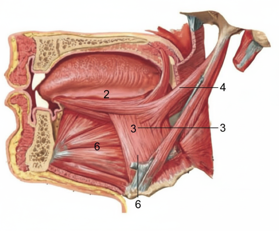
Identify the structure labeled as genioglossus in the given image showing a sagittal view of the oral cavity and pharynx.
The antagonist of superior rectus is:
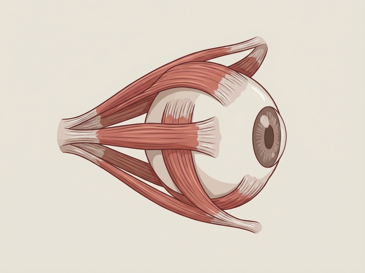
The image shows the lateral pterygoid muscle. What is the primary action of this muscle on the mandible?
Which of the following is correct about the image shown below?
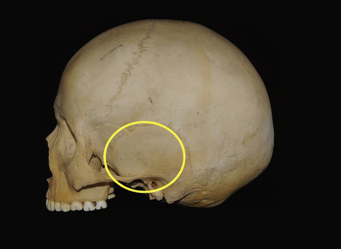
The highlighted part in the image is called
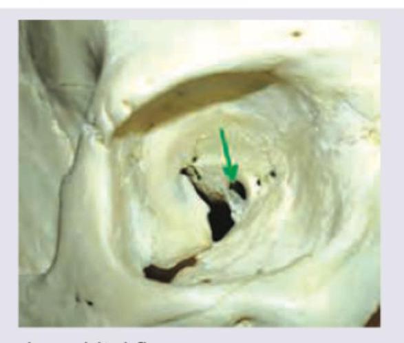
The image shows $\qquad$ (marked by green arrow):
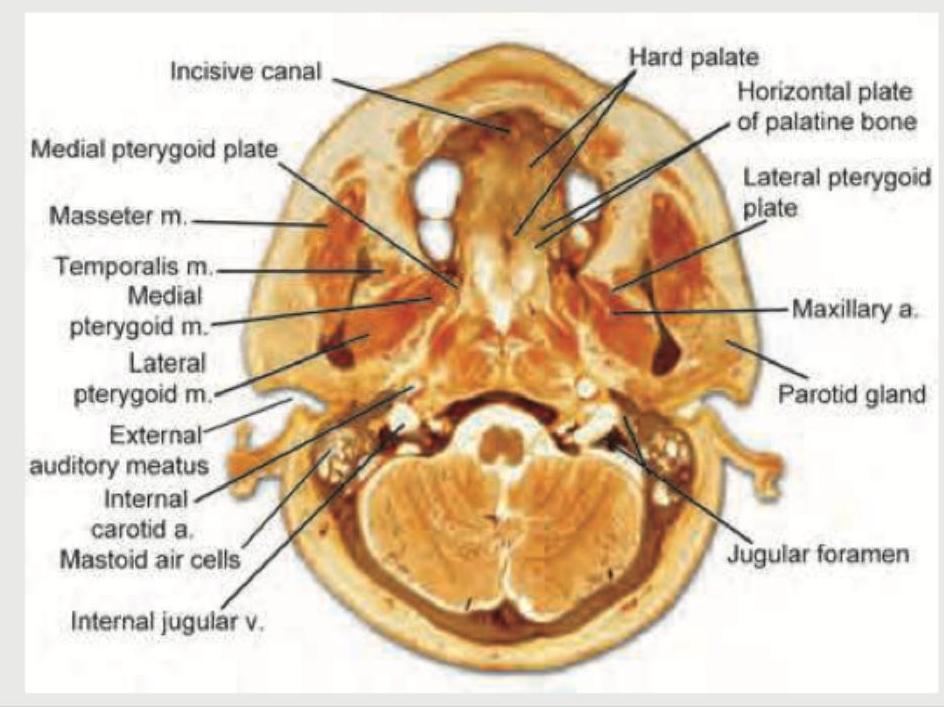
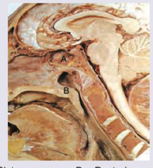
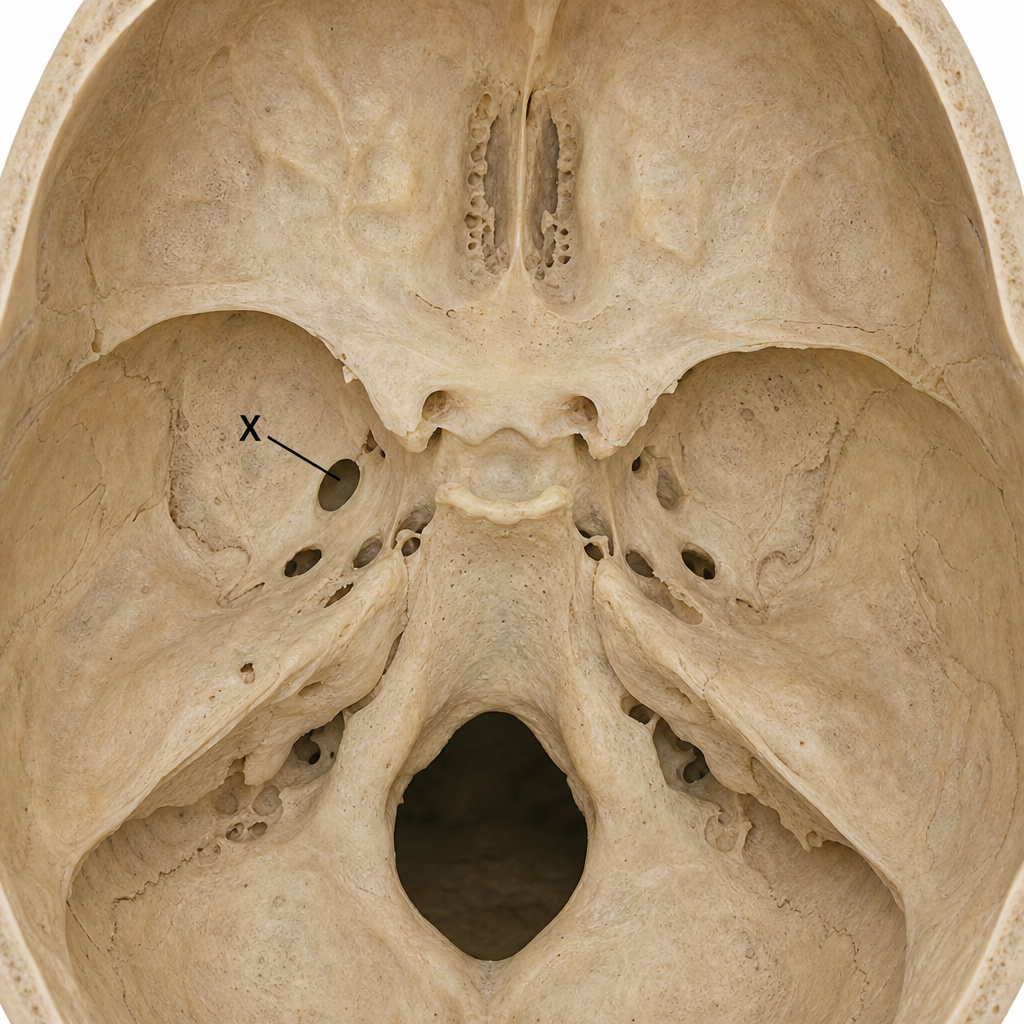
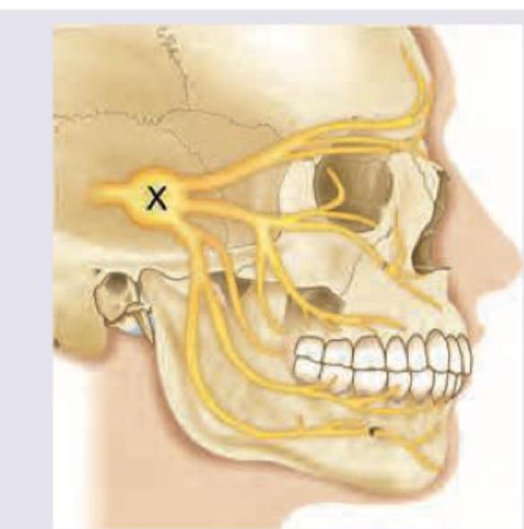
Identify the foramen marked below as $X$ :
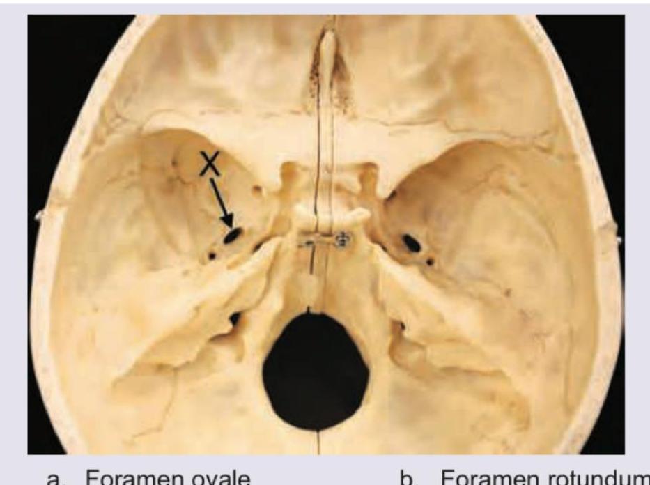
Identify the foramen marked below?
The secreto-motor fibers to the gland shown in the figure pass through?
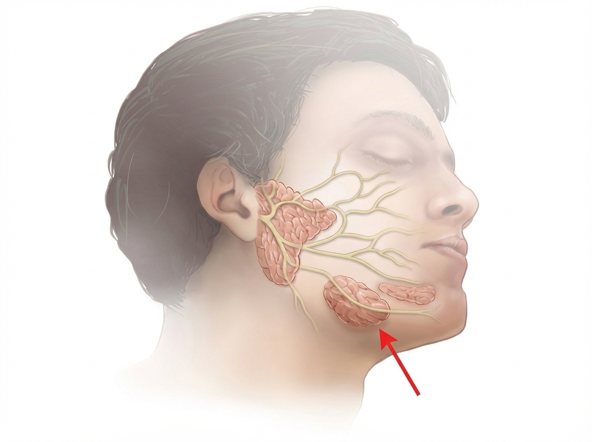
All are true about the ganglion ' $X$ ' shown in the image except:
Practice by Chapter
Skull and Facial Bones
Practice Questions
Scalp and Facial Muscles
Practice Questions
Dural Venous Sinuses
Practice Questions
Cranial Cavity
Practice Questions
Orbit and Contents
Practice Questions
Temporal and Infratemporal Regions
Practice Questions
Pterygopalatine Fossa
Practice Questions
Oral Cavity
Practice Questions
Paranasal Sinuses
Practice Questions
Applied Anatomy and Clinical Correlations
Practice Questions
Want unlimited practice?
Get full access to all questions, explanations, and performance tracking.
Scan to download app