
Head — MCQs

On this page
What overlies the lateral wall of the mastoid antrum?
The tendons of the Sartorius, gracilis, and semitendinosus muscles form the pes anserinus at the neck of the tibia. A similar type of structure is also seen in which of the following?
Which of the following is NOT a terminal branch of the facial nerve?
Which of the following structures is NOT supplied by the mandibular nerve?
The nasal septum is formed by all of the following structures EXCEPT:
Which muscle is primarily responsible for the opening of the mouth?
Which statement best describes the cranial fossae?
A patient presented to OPD with ophthalmoplegia and ptosis. Diagnosis of superior orbital fissure syndrome was confirmed after examination. Which nerves are compressed in this case ?
The upper part of the uncinate process commonly attaches to?
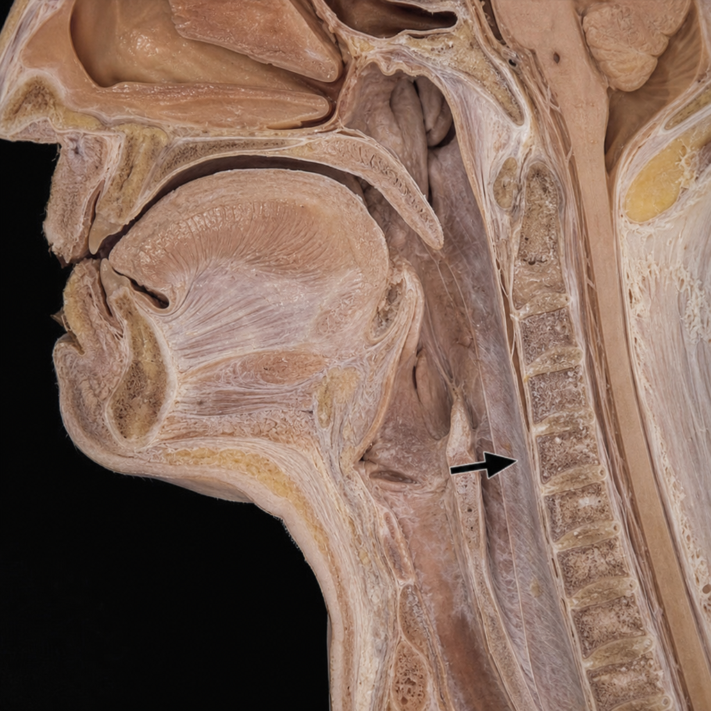
Identify the structure indicated by the arrow in the image.
Practice by Chapter
Skull and Facial Bones
Practice Questions
Scalp and Facial Muscles
Practice Questions
Dural Venous Sinuses
Practice Questions
Cranial Cavity
Practice Questions
Orbit and Contents
Practice Questions
Temporal and Infratemporal Regions
Practice Questions
Pterygopalatine Fossa
Practice Questions
Oral Cavity
Practice Questions
Paranasal Sinuses
Practice Questions
Applied Anatomy and Clinical Correlations
Practice Questions
Want unlimited practice?
Get full access to all questions, explanations, and performance tracking.
Scan to download app