
Head — MCQs

On this page
While giving an incision for third molar trans-alveolar extraction, what anatomical structure is at risk if the posterior extension of the incision is given in a straight line due to mandibular anatomy?
The nerve to the pterygoid canal is formed from which combination of nerves?
Which of the following is a secondary site of spread of odontogenic infection involving the pterygomandibular space?
Which cranial nerve does NOT carry taste sensation from the tongue?
Circumvallate papillae are present:
The ascending palatine artery is a branch of:
Which of the following statements about the lateral pterygoid muscle is true?
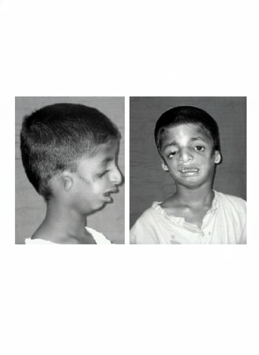
The image shows a patient with a congenital craniofacial condition. Which of the following is a striking feature of this condition?
A 54-year-old man presents with severe pain in his nasal cavity. Radiographic examination reveals a carcinoma in his nasal cavity. In which of the following locations would the carcinoma block the hiatus of the maxillary sinus?
When a patient's chin and mandible deviate to the right upon opening, which of the following is a possible cause?
Practice by Chapter
Skull and Facial Bones
Practice Questions
Scalp and Facial Muscles
Practice Questions
Dural Venous Sinuses
Practice Questions
Cranial Cavity
Practice Questions
Orbit and Contents
Practice Questions
Temporal and Infratemporal Regions
Practice Questions
Pterygopalatine Fossa
Practice Questions
Oral Cavity
Practice Questions
Paranasal Sinuses
Practice Questions
Applied Anatomy and Clinical Correlations
Practice Questions
Want unlimited practice?
Get full access to all questions, explanations, and performance tracking.
Scan to download app