
Head — MCQs

On this page
What is the largest paranasal sinus?
All of the following statements about Ebner glands are true, EXCEPT:
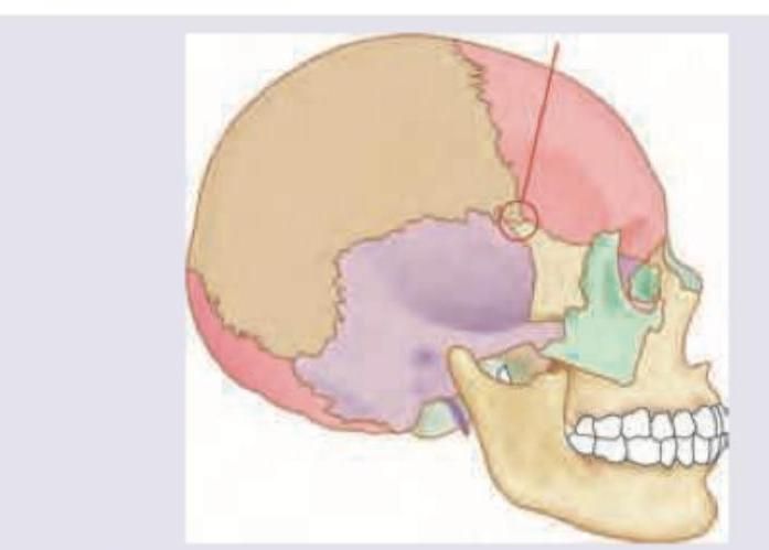
The marked area in the skull represents:
The deep surface of the hyoglossus muscle is related to which of the following structures?
Glands of Zeis are:
In cavernous sinus thrombosis, which is the first cranial nerve to be affected?
All of the following muscles are innervated by the facial nerve EXCEPT?
Which of the following statements is NOT true regarding the lacrimal gland?
What is the deepest layer of the scalp?
The anterior ethmoidal artery is closely related to which structure?
Practice by Chapter
Skull and Facial Bones
Practice Questions
Scalp and Facial Muscles
Practice Questions
Dural Venous Sinuses
Practice Questions
Cranial Cavity
Practice Questions
Orbit and Contents
Practice Questions
Temporal and Infratemporal Regions
Practice Questions
Pterygopalatine Fossa
Practice Questions
Oral Cavity
Practice Questions
Paranasal Sinuses
Practice Questions
Applied Anatomy and Clinical Correlations
Practice Questions
Want unlimited practice?
Get full access to all questions, explanations, and performance tracking.
Scan to download app