Consider the following statements with reference to scalp : 1. The blood vessels lie within dense connective tissue. 2. The anterior scalp is supplied by supraorbital and supratrochlear vessels. 3. The lateral and posterior scalp is supplied by superficial temporal, posterior auricular and occipital arteries. Which of the statements given above are correct?
Important landmark in submandibular gland dissection is:
Which one of the following statements regarding anatomy of fetal head is NOT true?
Cephalhematomas are most commonly found over:
A pregnant woman presents with fever, retroorbital pain, headache, pulsatile proptosis of the right eye, and tinnitus. BP and fundus were normal. Which of the following structures are involved?
What is the action of the superior oblique muscle?
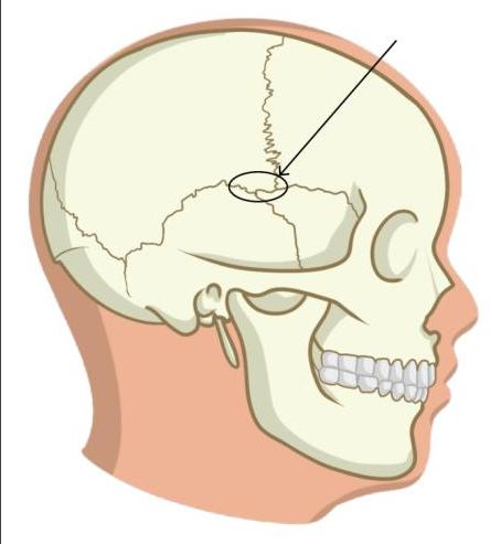
Identify the incorrect statement regarding the marked structure.

Masseter is supplied by which nerve?
Cutaneous supply over the parotid gland is by:
Which structure doesn't pass through the parotid gland?
Explanation: ***1, 2 and 3*** - All three statements provide accurate anatomical details regarding the scalp's structure and blood supply. The specific arrangement of vessels within the **dense connective tissue** and the listed arterial branches are correct. - The scalp is known for its rich vascularity and its five distinct layers, with the **dense connective tissue** layer anchoring the vessels, and a broad arterial supply covering all regions. *2 and 3 only* - This option is incorrect because statement 1, regarding the blood vessels lying within **dense connective tissue**, is also a correct anatomical fact. - Omitting statement 1 overlooks a crucial structural characteristic of the scalp layers, specifically the second layer (C for connective tissue). *1 and 3 only* - This option is incorrect because statement 2, detailing the supply of the anterior scalp by **supraorbital** and **supratrochlear vessels**, is also accurate. - Excluding statement 2 means ignoring a key component of the arterial supply to the anterior region of the scalp. *1 and 2 only* - This option is incorrect because statement 3, which describes the blood supply to the lateral and posterior scalp by the **superficial temporal**, **posterior auricular**, and **occipital arteries**, is also correct. - Failing to include statement 3 results in an incomplete description of the scalp's extensive and varied arterial network.
Explanation: ***Posterior border of mylohyoid muscle*** - Lifting the **posterior border of the mylohyoid muscle** exposes the deep part of the submandibular gland and crucial structures like the **lingual nerve** and **Wharton's duct**. - This landmark serves as a critical guide during dissection, helping to protect adjacent nerves and vessels while ensuring complete gland removal. *Anterior belly of digastric muscle* - The anterior belly of the digastric muscle forms the anterior boundary of the **digastric triangle**, which contains the superficial portion of the submandibular gland. - While relevant for defining the superficial borders, it is not the key landmark for accessing the deep part of the gland or protecting vital structures during dissection. *Facial artery* - The **facial artery** is closely associated with the submandibular gland, usually looping over or through it, but it is not a primary surgical landmark for deeper dissection or critical structure identification. - It is often ligated or preserved during dissection due to its proximity, but its position can vary, making it less reliable as a consistent access guide. *Posterior belly of digastric muscle* - The posterior belly of the digastric muscle forms the posterior boundary of the digastric triangle. - It is relevant for defining the superficial boundaries of the gland but does not provide direct access or guide the deeper dissection of the submandibular gland or protection of critical associated nerves.
Explanation: The lambdoid suture separates the two parietal bones - This statement is **incorrect**. The **sagittal suture** separates the two parietal bones [1]. - The **lambdoid suture** separates the parietal bones from the occipital bone [1]. *Frontal suture separates the two frontal bones* - This statement is **true**. The frontal suture (also called the metopic suture) divides the two halves of the **frontal bone** in the fetal skull [1]. - While it usually fuses after birth, it is prominent in the fetal head. *Bregma is a diamond shaped space at junction of coronal and sagittal sutures* - This statement is **true**. The **bregma** is the anterior fontanelle, a significant landmark in the fetal skull, formed by the junction of the coronal and sagittal sutures. - Its diamond shape and location are crucial for assessing fetal head position and molding during labor [2]. *Coronal suture separates frontal bones from parietal bones* - This statement is **true**. The **coronal suture** runs bilaterally across the top of the skull, separating the **frontal bone** anteriorly from the two **parietal bones** posteriorly [1]. - This anatomical arrangement allows for molding of the fetal head during passage through the birth canal [1].
Explanation: ***Parietal bone*** - The **parietal bones** are the most common site for cephalhematomas due to their prominence and susceptibility to trauma during vaginal delivery [1]. - The **force of uterine contractions** and contact with the birth canal can cause blood vessels beneath the periosteum of the parietal bone to rupture [1]. *Occipital bone* - While possible, cephalhematomas are less commonly observed over the **occipital bone** compared to the parietal region. - The **occipital protrusion** is less frequently subjected to the specific shearing forces that cause subperiosteal bleeding. *Temporal bone* - Cephalhematomas over the **temporal bone** are rare, primarily because this area is less frequently directly impacted during birth. - The relatively thinner bone and surrounding muscles also provide some protection against the typical trauma leading to this condition. *Frontal bone* - Cephalhematomas are generally uncommon over the **frontal bone**. - This area is less exposed to direct pressure and friction from the maternal pelvis during delivery.
Explanation: Abducens nerve, Oculomotor nerve, Internal carotid artery, Cavernous sinus - The combination of retroorbital pain, pulsatile proptosis, and tinnitus in a pregnant woman (indicating a potential hypercoagulable state) strongly suggests a carotid-cavernous fistula. - A carotid-cavernous fistula directly connects the high-pressure internal carotid artery to the low-pressure cavernous sinus, causing engorgement of the sinus and venous backflow. This can directly impact structures passing through or adjacent to the cavernous sinus, explaining the range of symptoms. - The oculomotor nerve (CN III) and abducens nerve (CN VI) both traverse the cavernous sinus and are commonly affected, leading to ophthalmoplegia and cranial nerve palsies. Abducens nerve, Oculomotor nerve, Internal carotid artery - This option incorrectly omits the cavernous sinus, which is central to the pathophysiology of the symptoms presented. - The cavernous sinus is the anatomical location where the internal carotid artery and multiple cranial nerves (including abducens and oculomotor) are in close proximity, making its involvement crucial for the observed signs. Oculomotor nerve, Internal carotid artery, Cavernous sinus - This option incorrectly omits the abducens nerve. While not explicitly stated as palsy, this nerve is often affected alongside the oculomotor nerve due to its passage through the cavernous sinus. - Involvement of the cavernous sinus often leads to dysfunction of cranial nerves III, IV, V1, V2, and VI, making abducens nerve involvement highly probable in such cases. Abducens nerve, Internal carotid artery, Cavernous sinus - This option incorrectly omits the oculomotor nerve. Pulsatile proptosis and retroorbital pain are strong indicators of cavernous sinus pathology, which frequently involves the oculomotor nerve (CN III), leading to ophthalmoplegia or partial palsies. - The oculomotor nerve is particularly vulnerable due to its course within the lateral wall of the cavernous sinus, making its involvement a common finding in cavernous sinus disorders.
Explanation: ***Intorsion, abduction, depression*** - The **superior oblique muscle** has three actions: **depression** (downward movement - primary action), **abduction** (movement away from the midline), and **intorsion** (medial/internal rotation of the globe) [1]. - The depressor action is most effective when the eye is **adducted** (turned inward), as the muscle's line of pull is then optimally aligned with the vertical axis. - Mnemonic: **SO-DAI** (Superior Oblique: Depression, Abduction, Intorsion) *Intorsion, adduction, elevation* - While **intorsion** is correct, this option incorrectly includes **adduction** and **elevation**. - The superior oblique causes **abduction** (not adduction) and **depression** (not elevation) [1]. - **Elevation** is performed by the superior rectus and inferior oblique muscles [1]. *Extorsion, adduction, elevation* - All three actions are incorrect for the superior oblique muscle. - The correct actions are **intorsion**, **abduction**, and **depression**. - **Extorsion** is performed by the inferior oblique and inferior rectus muscles [1]. *Extorsion, abduction, depression* - While **abduction** and **depression** are correct, **extorsion** is wrong. - The superior oblique causes **intorsion** (internal rotation), not extorsion (external rotation). - This is a common point of confusion - remember the superior oblique **intorts**, while the inferior oblique **extorts**.
Explanation: *It is the meeting point of frontal, parietal, temporal and sphenoid bones.* - The marked structure is the **pterion**, which is indeed the junction of the **frontal, parietal, temporal, and sphenoid bones**. - This statement is **correct**, as it accurately describes the anatomical composition of the pterion. ***Accessory middle meningeal artery lies under this structure*** - This is the **INCORRECT statement** and hence the correct answer. - The **middle meningeal artery** (not the accessory middle meningeal artery) runs in a groove deep to the pterion. - A fracture at the pterion can lacerate the **middle meningeal artery**, leading to an **epidural hematoma**. - The accessory middle meningeal artery is a separate vessel that does not typically lie under the pterion. *Blow to the lateral side of the skull injures the marked structure* - The pterion is the **thinnest part of the lateral wall of the skull**, making it vulnerable to fracture from a lateral blow. - Due to its thinness and underlying structures, trauma to this area is clinically significant. - This statement is **correct**. *It corresponds to site of anterolateral fontanelle of fetal skull* - The pterion in the adult skull corresponds to the former site of the **anterolateral (sphenoidal) fontanelle** in the fetal skull. - This fontanelle allows for skull molding during birth and brain growth postnatally. - This statement is **correct**.
Explanation: ***Mandibular*** - The **masseter muscle** is a muscle of mastication, and all muscles of mastication are exclusively innervated by the **mandibular division** (V3) of the trigeminal nerve. - The mandibular nerve carries both **motor and sensory fibers**; its motor branches supply the masseter, temporalis, medial and lateral pterygoids, mylohyoid, anterior belly of digastric, tensor veli palatini, and tensor tympani. *Glossopharyngeal* - The **glossopharyngeal nerve (CN IX)** primarily supplies sensation to the posterior third of the tongue, the pharynx, and the tympanic membrane. - It also has motor functions, innervating the **stylopharyngeus muscle** and providing parasympathetic innervation to the parotid gland. *Facial* - The **facial nerve (CN VII)** is primarily responsible for innervating the **muscles of facial expression**. - It also carries taste sensation from the anterior two-thirds of the tongue and provides parasympathetic innervation to the lacrimal, submandibular, and sublingual glands. *Hypoglossal* - The **hypoglossal nerve (CN XII)** is exclusively a motor nerve. - It innervates all of the **intrinsic and extrinsic muscles of the tongue**, except for the palatoglossus muscle (which is innervated by the vagus nerve).
Explanation: ***Greater auricular nerve*** - The **greater auricular nerve**, a branch of the cervical plexus (C2-C3), is the **primary cutaneous nerve** supplying sensation to the skin over the parotid gland and the angle of the mandible. - It ascends from behind the sternocleidomastoid muscle and provides the main sensory innervation to the parotid region, making it the most important nerve for this area. *Auriculotemporal nerve* - The **auriculotemporal nerve** (branch of mandibular division of trigeminal nerve) supplies sensory innervation to the skin of the temporal region, the external auditory meatus, and the tympanic membrane. - While it provides some cutaneous supply to the upper/posterior part of the parotid region, the **greater auricular nerve is the primary cutaneous nerve** over the parotid gland proper. - It also carries postganglionic parasympathetic fibers (from otic ganglion) to the parotid gland for secretomotor function. *Greater occipital nerve* - The **greater occipital nerve** is responsible for cutaneous sensation to the posterior scalp, up to the vertex of the head. - It arises from the dorsal ramus of C2 and has no role in the cutaneous supply over the parotid gland. *Facial nerve* - The **facial nerve (CN VII)** is primarily a motor nerve that controls the muscles of facial expression. - While it passes through the parotid gland and divides within it, it does not provide cutaneous sensory innervation to the skin overlying the gland.
Explanation: ***Internal Carotid Artery*** - The **internal carotid artery** does not pass through the parotid gland; it is located medial to the gland. - It ascends in the neck to supply the brain and does not traverse the glandular parenchyma. *Retromandibular vein* - The retromandibular vein is formed within the parotid gland by the union of the **superficial temporal vein** and the **maxillary vein**. - It then exits the gland and divides into anterior and posterior divisions. *Facial nerve* - The **facial nerve (cranial nerve VII)** enters the posterior aspect of the parotid gland and divides into its five terminal branches within the gland. - It supplies motor innervation to the **muscles of facial expression**. *External Carotid Artery and its branches* - The **external carotid artery** passes through the parotid gland, where it gives off several important branches. - The main branches within the gland are the **maxillary artery** and the **superficial temporal artery**.
Skull and Facial Bones
Practice Questions
Scalp and Facial Muscles
Practice Questions
Dural Venous Sinuses
Practice Questions
Cranial Cavity
Practice Questions
Orbit and Contents
Practice Questions
Temporal and Infratemporal Regions
Practice Questions
Pterygopalatine Fossa
Practice Questions
Oral Cavity
Practice Questions
Paranasal Sinuses
Practice Questions
Applied Anatomy and Clinical Correlations
Practice Questions
Get full access to all questions, explanations, and performance tracking.
Start For Free