Match the following 1. Hirschsprung's disease 2. Posterior urethral valve 3. Choledochal cyst 4. Intussusception A. Jaundice B. Currant jelly stools C. Distended abdomen D. Oligohydramnios
A 3-month-old with projectile vomiting and olive-shaped mass in abdomen is diagnosed with?
The prostate gland is derived from?
A patient presents with fecal discharge from the umbilicus. What is the most likely diagnosis?
Tenesmus occurs in lesions of the colon.
The labia majora develop from which embryological structure?
Congenital megacolon is confirmed by:
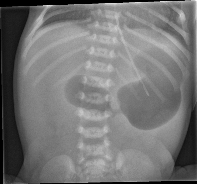
Omphalocele is caused by:-
All of the following organs develop in the mesentery of the stomach except for which of the following?
Embryologically, from which structure is the cerebellum derived?

Explanation: ***Correct Answer: 1-C, 2-D, 3-A, 4-B*** **Correct Associations:** - **Hirschsprung's disease (1) → Distended abdomen (C)**: Congenital absence of ganglion cells in the distal bowel leads to functional obstruction and subsequent abdominal distension. This is a hallmark presentation in neonates and infants. - **Posterior urethral valve (2) → Oligohydramnios (D)**: Urethral obstruction in utero prevents normal fetal urine output, resulting in decreased amniotic fluid (oligohydramnios). This can be detected on prenatal ultrasound. - **Choledochal cyst (3) → Jaundice (A)**: Congenital dilatation of the bile ducts causes biliary obstruction, presenting with jaundice as part of the classic triad (jaundice, abdominal mass, and pain). - **Intussusception (4) → Currant jelly stools (B)**: Telescoping of bowel causes mucosal ischemia and venous congestion, leading to bloody mucoid stools with characteristic "currant jelly" appearance. This is a pathognomonic feature. *Incorrect: 1-C, 2-D, 3-B, 4-A* - Incorrectly associates choledochal cyst with currant jelly stools (which is specific to intussusception) and intussusception with jaundice (which indicates biliary pathology). *Incorrect: 1-A, 2-D, 3-B, 4-C* - Wrongly links Hirschsprung's disease with jaundice instead of its characteristic abdominal distension, and misidentifies intussusception's primary feature. *Incorrect: 1-D, 2-C, 3-A, 4-B* - Swaps the associations between Hirschsprung's disease and PUV. Oligohydramnios is specific to urinary tract obstruction (PUV), not intestinal pathology (Hirschsprung's).
Explanation: ***Pyloric stenosis*** - The classic triad of **projectile vomiting**, a palpable **olive-shaped mass** (hypertrophied pylorus), and age of presentation (2-8 weeks, though 3 months is still possible) are highly indicative of **pyloric stenosis**. - This condition involves thickening of the **pyloric muscle**, leading to gastric outlet obstruction and non-bilious emesis. *Hirschsprung disease* - This typically presents with **constipation**, **abdominal distension**, and failure to pass meconium, rather than projectile vomiting. - It results from the absence of **ganglion cells** in the distal colon, causing functional obstruction. *GERD* - While GERD can cause vomiting in infants, it is usually not **projectile** and is rarely associated with a palpable **olive-shaped mass**. - Infants with GERD typically respond to conservative measures like thickening feeds or acid suppressants. *Duodenal atresia* - This condition presents with **bilious vomiting** (as the obstruction is distal to the ampulla of Vater) usually within the first 24-48 hours of life. - An abdominal X-ray would show a **double bubble sign**, which is not mentioned in the presentation for pyloric stenosis.
Explanation: ***Urogenital sinus*** - The **prostate gland** develops from endodermal buds that arise from the **urethral epithelium** of the urogenital sinus during the third month of gestation. - These buds grow into the surrounding mesenchyme, which differentiates into the stromal and smooth muscle components of the prostate. *Urogenital folds* - The **urogenital folds** (or urethral folds) in males fuse to form the **spongy urethra** and the ventral aspect of the penis. - In females, these folds remain separate and form the labia minora. *Labioscrotal swelling* - The **labioscrotal swellings** in males fuse in the midline to form the **scrotum**. - In females, these swellings remain unfused and give rise to the labia majora. *Gubernaculum* - The **gubernaculum** is a mesenchymal cord that plays a crucial role in the **descent of the testes** into the scrotum. - It guides the migrating testis and anchors it to the scrotal floor.
Explanation: ***Patent vitelline duct*** - **Fecal discharge from the umbilicus** indicates a persistent communication between the **ileum** and the **umbilicus** through a patent vitelline (omphalomesenteric) duct. - This congenital anomaly represents a remnant of the **omphalomesenteric duct** that **completely failed to involute**, creating a **fistulous tract** allowing intestinal contents to exit through the umbilicus. - This is the **most complete form** of vitelline duct persistence (other forms include Meckel's diverticulum, fibrous band, or umbilical polyp). *Urachal fistula* - A urachal fistula occurs when the **urachus** remains patent, creating a connection between the **bladder** and the umbilicus. - While it can result in umbilical discharge, the discharge would be **urine**, not feces. *Omphalocele* - An omphalocele is a **congenital abdominal wall defect** where abdominal contents protrude into a sac at the base of the umbilicus. - It does not involve a fistulous communication with intestines causing fecal discharge, but rather a **herniation** of organs covered by a peritoneal membrane. *Gastroschisis* - Gastroschisis is a congenital anomaly characterized by the **protrusion of abdominal organs** directly into the amniotic cavity **without a covering sac**, usually to the **right of the umbilicus**. - Like omphalocele, it's a **herniation defect** presenting at birth and does not involve an abnormal fistulous connection causing fecal discharge from the umbilicus.
Explanation: ***Sigmoid colon*** - Lesions in the **sigmoid colon** are most commonly associated with **tenesmus** because of its proximity to the rectum and its role in stool storage and defecation. - Inflammation or obstruction in this region can trigger a constant urge to defecate, even when the bowel is empty. *Lesions on the right side of the colon* - **Right-sided colon lesions** are typically associated with symptoms like **anemia**, occult bleeding, and vague abdominal pain due to their larger luminal diameter, rather than tenesmus. - The right colon is primarily involved in **water and electrolyte absorption**, and lesions here don't usually cause the sensation of incomplete evacuation linked to tenesmus. *Lesions in the descending colon* - While the **descending colon** is involved in stool transit, lesions here are less likely to cause prominent tenesmus compared to the sigmoid colon. - Symptoms usually involve **changes in bowel habits**, abdominal pain, or blood in stool, but typically without the persistent urgency characteristic of tenesmus. *Ileal lesions* - **Ileal lesions** (in the small intestine) are associated with symptoms like malabsorption, abdominal pain, and diarrhea [2]. - The ileum is located far from the rectum and anal canal, so it does not directly influence the sensation of **incomplete defecation** or tenesmus [1].
Explanation: ***Labioscrotal swellings*** - The **labia majora** develop from the **labioscrotal swellings**, which are paired bilateral structures that appear around week 9-10 of development [1]. - These swellings arise lateral to the urogenital folds and do not fuse in females, forming the labia majora. - In males, these same structures fuse in the midline to form the scrotum. - This is a key example of **sexual differentiation** in embryological development [1]. *Urogenital folds* - The urogenital folds form the **labia minora** in females, not the labia majora. - In males, these folds fuse to form the ventral aspect of the penis and enclose the penile urethra. *Genital tubercle* - The genital tubercle forms the **clitoris** in females and the **glans penis** in males. - It does not contribute to the formation of the labia majora. *Müllerian ducts* - The Müllerian (paramesonephric) ducts form the **upper vagina, uterus, and fallopian tubes** in females. - They are internal structures and do not contribute to external genitalia like the labia majora.
Explanation: ***Rectal biopsy*** - **Rectal biopsy** is the **gold standard** for diagnosing congenital megacolon (Hirschsprung disease) by demonstrating the **absence of ganglion cells** in the myenteric and submucosal plexuses [1]. - This absence of innervation leads to a functional obstruction, causing proximal bowel dilation [1]. *Sigmoidoscopy* - While **sigmoidoscopy** allows visualization of the mucosa, it cannot directly confirm the **absence of ganglion cells**, which is the hallmark of Hirschsprung disease. - It may reveal features like a **narrowed segment** transition to a dilated segment but requires further diagnostic confirmation. *Invertogram* - An **invertogram** is used to assess the **anal position** and check for imperforate anus by showing the gas bubble in the rectum relative to the skin marker. - It does not provide information about the **innervation of the rectosigmoid colon** or the presence of ganglion cells. *Barium enema* - A **barium enema** can reveal characteristic findings like a **transition zone** between a narrowed aganglionic segment and a dilated, normally innervated colon. - However, it is a **radiological study** and cannot definitively confirm the histopathological absence of ganglion cells, which requires a tissue biopsy. **References:** [1] Kumar V, Abbas AK, et al.. Robbins and Cotran Pathologic Basis of Disease. 9th ed. The Gastrointestinal Tract, pp. 759-760.
Explanation: Failure of gut to return to body cavity after its physiological herniation - **Omphalocele** results from a failure of the midgut to properly return to the abdominal cavity after its normal physiological herniation into the umbilical cord during fetal development (around the 6th to 10th week of gestation). - The abdominal muscles fail to close, and the herniated organs (intestines, liver) are covered by a **peritoneal sac** at the base of the umbilical cord. *Duplication of intestinal loops* - **Intestinal duplications** are congenital malformations typically presenting as cystic or tubular structures that are intimately associated with the gastrointestinal tract. [1] - They do not involve the herniation of abdominal contents outside the body cavity. *Abnormal rotation of intestinal loops* - **Malrotation of the gut** refers to abnormal positioning of the intestines within the abdominal cavity due to incomplete or arrested rotation during development. - This can lead to conditions like volvulus but does not involve the organs being outside the abdominal cavity. *Reversed rotation of intestinal loops* - **Reversed rotation** is a very rare form of malrotation where the third stage of midgut rotation occurs in reverse, leading to the small intestine being positioned anterior to the superior mesenteric artery and the colon posterior. - This condition affects the internal arrangement of organs within the abdomen, not their external herniation.
Explanation: ***Kidney*** - The **kidneys** develop in the **retroperitoneal space** [2], external to the peritoneal cavity and not within the mesentery of the stomach. - Their development is distinct from the foregut derivatives and associated mesenteries. *Liver and gallbladder* - The **liver** and **gallbladder** develop as outgrowths from the **foregut endoderm** within the ventral mesentery of the stomach [1]. - They remain connected to the stomach by the lesser omentum, which is derived from the ventral mesentery [1]. *Spleen and pancreas* - The **spleen** develops within the **dorsal mesentery** of the stomach. - The **pancreas** develops from both ventral and dorsal buds; the dorsal bud forms in the **dorsal mesentery** and the ventral bud in the **ventral mesentery** [1]. *None of the options* - This option is incorrect because the **kidney** is the correct answer; it does not develop from the mesentery of the stomach.
Explanation: ***Metencephalon*** - The cerebellum develops from the **metencephalon**, a secondary brain vesicle that arises from the **rhombencephalon** during embryonic development. - This structure is responsible for coordinating muscle movements and balancing, which are key functions of the **cerebellum**. *Prosencephalon* - The **prosencephalon** forms the **forebrain**, which includes structures such as the **telencephalon** and **diencephalon**, not the cerebellum. - Its derivatives primarily relate to higher cognitive functions and sensory processing rather than motor control. *Mesencephalon* - The **mesencephalon** develops into the **midbrain**, which is involved in visual and auditory processing, but does not contribute to the formation of the cerebellum. - It also plays a role in motor control via connections with other brain regions, yet lacks the direct association with the cerebellar structure. *Rhombencephalon* - The **rhombencephalon** is a primary brain vesicle that gives rise to both the **metencephalon** and **myelencephalon**, but is not the final structure of the cerebellum itself. - While it lays the groundwork for the hindbrain, the cerebellum specifically originates from the **metencephalon** within this region.
Get full access to all questions, explanations, and performance tracking.
Start For Free