Sumoylation of histone proteins is associated with
Phenotypic expression of a gene depending on the parent of origin is referred to as:
The following gene mutation protects tumor cells from apoptosis:
Gene not involved in SCID:
Which of the following protein molecules is responsible for cell-to-cell adhesion?
Which of the following is not a part of extracellular matrix (ECM)?
At what week of gestation do limb buds appear?
The following ocular structure is not derived from surface ectoderm –
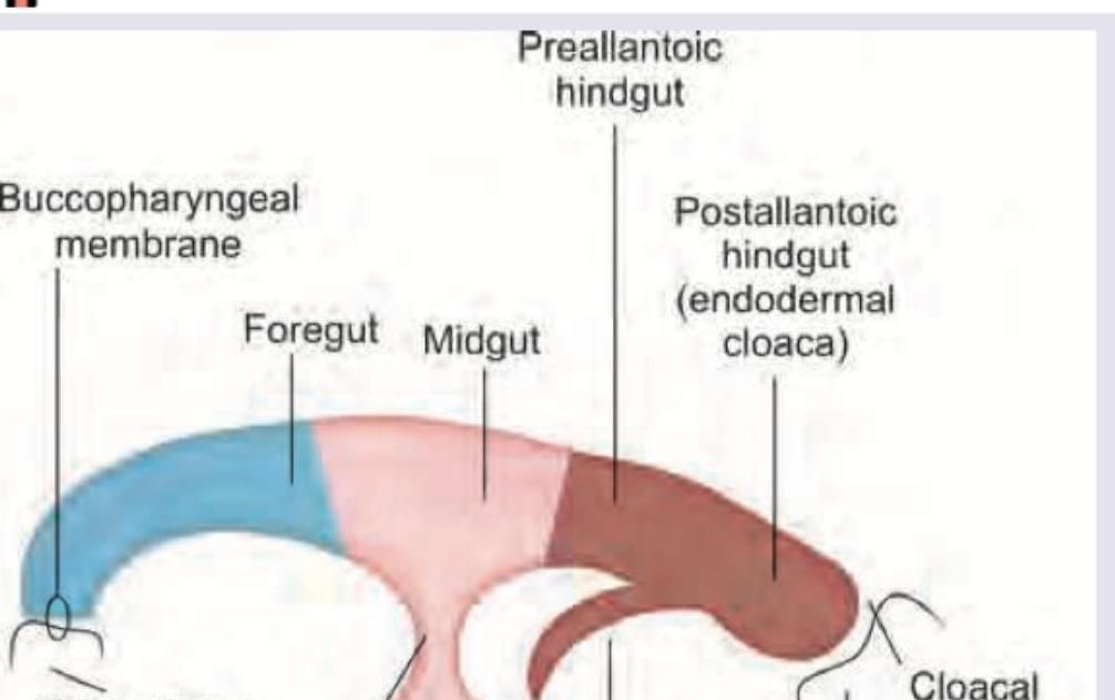
Which is wrong about the image given below?

Which paranasal sinuses are present at birth?
Explanation: ***Transcription repression*** - **Sumoylation** is a post-translational modification involving the covalent attachment of **Small Ubiquitin-like Modifier (SUMO) proteins** to target proteins, which leads to transcriptional repression. - When histones are sumoylated, it alters chromatin structure and recruits **transcriptional corepressors**, making the DNA less accessible for transcription factors. - This is the **primary and well-established function** of histone sumoylation in gene regulation. *Activation of gene transcription* - **Histone acetylation** and specific methylation patterns (e.g., H3K4me3, H3K36me3) are associated with **transcriptional activation**, not sumoylation. - Sumoylation typically creates a repressive chromatin environment, hindering gene expression. *Condensation of chromosome* - While sumoylation can influence chromatin structure, **chromosome condensation** during cell division is primarily regulated by **condensins** and **cohesins**. - Sumoylation's role in condensation is indirect and not its primary function. *DNA replication* - DNA replication is a separate process from transcriptional regulation and involves DNA polymerases and replication machinery. - Histone sumoylation specifically affects **gene transcription**, not DNA replication.
Explanation: ***Genomic imprinting (parent-of-origin gene expression)*** - **Genomic imprinting** is an epigenetic phenomenon where gene expression is dependent on whether the gene was inherited from the mother or the father. - This results in monoallelic expression of specific genes, with only one copy (maternal or paternal) being active. *Mosaic genetic variation* - **Mosaicism** refers to the presence of two or more populations of genetically different cells in one individual, all derived from a single zygote. - This typically arises from a somatic mutation during development, not from differential expression based on parental origin. *Nonpenetrance of genotype* - **Nonpenetrance** occurs when individuals carrying a disease-causing genotype do not express the associated phenotype. - This concept relates to the presence or absence of a phenotype, not the differential expression based on parental origin. *Genetic anticipation* - **Genetic anticipation** is the phenomenon where the symptoms of a genetic disorder become more severe and/or appear at an earlier age in successive generations. - This is commonly observed in disorders caused by expansions of trinucleotide repeats, such as Huntington's disease, and is distinct from parent-of-origin gene expression.
Explanation: ***bcl-2*** - The **bcl-2 gene** produces a protein that inhibits apoptosis, thereby allowing tumor cells to evade programmed cell death [1][2]. - Overexpression of **bcl-2** is associated with various cancers, making it pivotal in cancer biology [1]. *RB* - The **RB gene** is primarily a tumor suppressor, regulating the cell cycle, and does not directly prevent apoptosis. - Loss of RB function leads to unregulated cell division rather than inhibition of cell death. *TGFβ* - **TGFβ** acts as a tumor suppressor and can induce apoptosis in certain contexts, particularly in oncogenic processes. - Its primary role involves regulating cell growth and differentiation, not directly protecting against apoptosis. *BRCA* - **BRCA genes** (BRCA1 and BRCA2) are involved in DNA repair mechanisms; mutations increase cancer susceptibility but do not prevent apoptosis directly. - Dysfunction in BRCA proteins primarily impacts the repair of DNA damage, leading to genomic instability rather than apoptosis resistance. **References:** [1] Kumar V, Abbas AK, et al.. Robbins and Cotran Pathologic Basis of Disease. 9th ed. Neoplasia, pp. 310-311. [2] Kumar V, Abbas AK, et al.. Robbins and Cotran Pathologic Basis of Disease. 9th ed. Neoplasia, p. 310.
Explanation: ***BTK*** - **Bruton's tyrosine kinase (BTK)** is associated with **X-linked agammaglobulinemia (XLA)**, a primary immunodeficiency characterized by the absence of mature B cells and significantly reduced antibody production. While it causes severe immune deficiency, it is not a direct cause of **SCID**. - XLA results in recurrent bacterial infections due to an inability to produce antibodies, rather than the severe combined T and B cell dysfunction seen in SCID. *ZAP70* - **ZAP70** deficiency is a cause of **SCID**. It leads to impaired T-cell receptor signaling, resulting in profound functional T-cell lymphopenia. - Patients with ZAP70 deficiency have normal numbers of CD4 T cells but very low or absent CD8 T cells, and their T cells are functionally impaired, leading to severe immunodeficiency. *IL2RG* - The **IL2RG** gene encodes the common gamma chain (γc), a crucial component of several **interleukin receptors (IL-2, IL-4, IL-7, IL-9, IL-15, IL-21)**. [1] - Mutations in IL2RG cause **X-linked SCID (X-SCID)**, the most common form of SCID, leading to a block in T-cell and NK-cell development due to defective cytokine signaling. [1] *JAK3* - **Janus kinase 3 (JAK3)** is a tyrosine kinase that associates with the **common gamma chain (γc)** and is essential for cytokine signaling downstream of the γc-containing receptors. [1] - **JAK3 deficiency** results in an **autosomal recessive form of SCID**, clinically indistinguishable from X-SCID, with impaired T-cell and NK-cell development due to defective cytokine signaling. [1] **References:** [1] Kumar V, Abbas AK, et al.. Robbins and Cotran Pathologic Basis of Disease. 9th ed. Diseases of the Immune System, pp. 247-248.
Explanation: ***Cadherin*** - **Cadherins** are transmembrane proteins that mediate **direct cell-to-cell adhesion** in a calcium-dependent manner - They form **adherens junctions** and **desmosomes**, which are essential for maintaining tissue integrity - Cadherins on adjacent cells bind to each other (**homophilic binding**), creating strong cell-cell connections - Critical for **embryonic development**, tissue architecture, and **epithelial barrier function** *Fibronectin* - **Fibronectin** is an extracellular matrix glycoprotein that mediates **cell-to-ECM adhesion**, not direct cell-to-cell adhesion - It binds to **integrins** on the cell surface, facilitating cell attachment to the extracellular matrix - Important for cell migration, wound healing, and embryonic development - Does not directly connect cells to each other *Collagen* - **Collagen** is the most abundant structural protein providing **tensile strength** to connective tissues - Primarily functions as **extracellular scaffolding**, not as an adhesion molecule - Provides mechanical support but does not mediate cell-cell adhesion *Laminin* - **Laminins** are major components of the **basal lamina** (basement membrane) - Mediate **cell-to-basal lamina adhesion** through integrin receptors - Important for cell differentiation, migration, and tissue organization - Function in cell-to-ECM adhesion, not cell-to-cell adhesion
Explanation: ***Lectins*** - **Lectins** are carbohydrate-binding proteins involved in various cellular processes but are typically found **on cell surfaces** or within cells, not as a major structural component of the ECM. - While they can interact with ECM components, they are not considered a direct structural element of the extracellular matrix itself. *Fibronectin* - **Fibronectin** is a critical **glycoprotein** in the ECM, playing a vital role in cell adhesion, growth, migration, and differentiation. - It links cells to collagen fibers and other ECM components, forming an essential scaffold. *Laminin* - **Laminin** is a major **glycoprotein** component of the **basal lamina**, a specialized layer of the ECM found beneath epithelial cells. - It helps in cell attachment, differentiation, and migration. *Proteoglycans* - **Proteoglycans** are macromolecules consisting of a **core protein** covalently linked to one or more **glycosaminoglycan (GAG) chains**. - They are abundant in the ECM, where they contribute to its structural integrity, hydration, and can regulate the diffusion of molecules.
Explanation: ***Week 4*** - The **upper limb buds** appear at the beginning of the fourth week, followed shortly by the **lower limb buds**. - This marks the crucial initial stage of **limb development** as mesenchymal outgrowths from the ventrolateral body wall. *Week 3* - This is the period of **gastrulation** and early **neurulation**, where the three germ layers are established and the neural tube begins to form. - While significant developmental events occur, the formation of visible **limb buds** has not yet begun. *Week 6* - By week 6, the limb buds have not only appeared but have undergone considerable development, with **hand and foot plates** becoming distinct. - The upper and lower limbs are beginning to show more defined structures, including the appearance of **digital rays**. *Week 9* - By week 9, the limbs are well-developed, with all major segments and **digits clearly visible**. - This stage is characterized by ongoing **ossification** and refined anatomical structures.
Explanation: **Sclera** - The **sclera** develops from the **neural crest cells**, which differentiate into mesenchymal tissue around the optic cup, forming the fibrous coats of the eye [1]. - It is part of the **fibrous tunic** of the eye, along with the cornea, and provides structural support. *Epithelium of lacrimal glands* - The **epithelium of lacrimal glands** originates from the **surface ectoderm** through invaginations and subsequent differentiation. - These glands are responsible for producing the **watery component of tears**. *Crystalline lens* - The **crystalline lens** also develops from the **surface ectoderm**, specifically from the lens placode, which invaginates to form the lens vesicle. - It is crucial for **focusing light** onto the retina. *Corneal epithelium* - The **corneal epithelium** is derived from the **surface ectoderm** and forms the outermost layer of the cornea [1]. - It provides a **protective barrier** and helps maintain the smooth refractive surface of the cornea [1].
Explanation: ***Dorsal mesentery degenerates*** - The **dorsal mesentery** persists and develops into various mesenteries supporting the abdominal organs (e.g., mesentery proper, transverse mesocolon, sigmoid mesocolon). It does not degenerate. - The **ventral mesentery degenerates** in areas due to the rotation of the gut and fusion processes, but the dorsal mesentery generally remains. *Foregut meets the stomodeum at bucco-pharyngeal membrane which ruptures at 4 weeks* - The **buccopharyngeal membrane separates the foregut from the stomodeum** (primitive mouth). - This membrane normally **ruptures around the 4th week** of development to establish communication between the oral cavity and the foregut. *Hindgut meets the proctodeum which ruptures at 8th week* - The **hindgut meets the proctodeum** (primitive anal pit) at the **cloacal membrane**. - This **cloacal membrane ruptures around the 7th or 8th week** of development, forming the anal opening. *Lining epithelium of gut is endodermal* - The **endoderm is the primary germ layer** that forms the lining epithelium of the entire gastrointestinal tract. - This includes the epithelium of the esophagus, stomach, intestines, and associated glands.
Explanation: The development of paranasal sinuses is a high-yield topic in embryology. At birth, only the **maxillary** and **ethmoid** sinuses are present and pneumatized enough to be clinically identifiable, though they are rudimentary in size. 1. **Maxillary Sinus:** This is the first sinus to develop (around the 3rd month of fetal life). At birth, it is a small sac measuring approximately 7 x 4 x 4 mm. 2. **Ethmoid Sinus:** These air cells are present at birth and continue to enlarge during early childhood. **Analysis of Incorrect Options:** * **Frontal Sinus:** This sinus is **absent at birth**. It starts developing from the anterior ethmoidal cells around the age of 2 and is usually not radiologically visible until age 5–7. * **Sphenoid Sinus:** While a tiny evagination may exist at birth, it is effectively **absent/non-pneumatized**. It begins to invade the sphenoid bone around age 3 and reaches full development in adolescence. **High-Yield Clinical Pearls for NEET-PG:** * **Sequence of Development:** Maxillary → Ethmoid → Sphenoid → Frontal (Mnemonic: **M**y **E**xtra **S**pecial **F**riend). * **Radiology:** The frontal sinus is the last to appear on an X-ray. * **Clinical Correlation:** Because the frontal and sphenoid sinuses are absent at birth, sinusitis in infants typically only involves the ethmoid or maxillary sinuses. * **Growth:** The maxillary sinus shows a rapid growth spurt during the eruption of permanent teeth.
Get full access to all questions, explanations, and performance tracking.
Start For Free