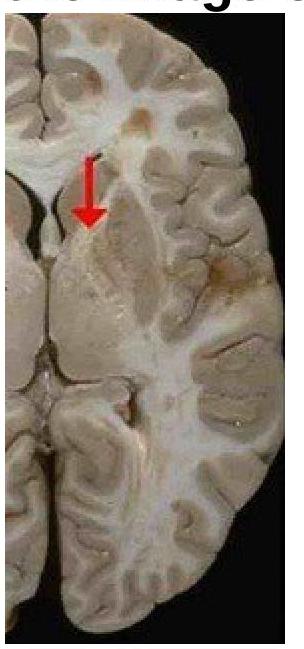
Identify the type of the fibre marked in the image of the internal capsule.

Where is the auditory cortex primarily located in the brain?
Which Brodmann's area is primarily associated with motor speech?
Superficial middle cerebral vein drains into -
Which nucleus is not seen in floor of the 4th ventricle -
The Great cerebral vein of Galen drains into which structure?
The third ventricle is a cavity located in which part of the brain?
Which tract is responsible for the ventral tegmental decussation in the cerebral peduncle?
Which thalamic nucleus has the most extensive reciprocal connections with the association areas of the neocortex?
Vertebral arteries of both sides unite to form
Explanation: ***Projection fibers*** - The image shows the **internal capsule**, which is a white matter structure composed of **projection fibers** that connect the cerebral cortex to subcortical structures, brainstem, and spinal cord. - These fibers facilitate communication between different levels of the central nervous system, including motor and sensory pathways. *Short association fibers* - These fibers, also known as **U-fibers**, connect adjacent gyri within the **same cerebral hemisphere**. - They are typically located superficially in the cerebral cortex, not deep within the brain as shown in the internal capsule. *Long association fibers* - These fibers connect **different lobes** within the **same cerebral hemisphere**, such as the arcuate fasciculus connecting temporal and frontal lobes. - While they are white matter tracts, they do not constitute the internal capsule, which is specifically known for its extensive projection pathways. *Commissural fibers* - **Commissural fibers** connect corresponding areas in the **two cerebral hemispheres**, with the most prominent example being the **corpus callosum**. - The internal capsule, shown in the image, primarily consists of fibers projecting superiorly and inferiorly, rather than horizontally across hemispheres.
Explanation: ***Superior temporal gyrus*** - The **primary auditory cortex** (Brodmann areas 41 and 42) is located in the **superior temporal gyrus**, primarily within the **transverse temporal gyri of Heschl**. [1] - This region is responsible for processing **auditory information**, including pitch, loudness, and sound localization. [1] *Inferior temporal gyrus* - The **inferior temporal gyrus** is a part of the temporal lobe involved in higher-level **visual processing** and object recognition. - It plays a role in the "what" pathway of vision and **memory formation**, not primary auditory processing. *Area 3,1,2* - **Brodmann areas 3, 1, and 2** collectively form the **primary somatosensory cortex**. [2] - This area is located in the **postcentral gyrus** of the parietal lobe and is responsible for processing touch, pain, temperature, and proprioception. [2] *Cingulate gyrus* - The **cingulate gyrus** is a component of the **limbic system**, involved in emotion formation, learning, memory, and executive function. - It plays a role in processing emotional aspects of pain and fear, but not primary auditory perception.
Explanation: Area 44 - **Brodmann Area 44** is primarily known as **Broca's area**, which is critical for **motor speech production** and language processing [1]. - Damage to this area typically results in **Broca's aphasia**, characterized by non-fluent speech and difficulty forming complete sentences [1]. Area 1, 2, 3 - These Brodmann areas constitute the **primary somatosensory cortex**, responsible for processing **tactile and proprioceptive information** from the body. - They are involved in sensory perception, not directly with motor speech production. Area 4, 6 - **Brodmann Area 4** is the **primary motor cortex**, involved in executing voluntary movements [2]. **Brodmann Area 6** is the **premotor and supplementary motor cortex**, involved in planning and coordinating movements [2]. - While these areas are crucial for motor control, they are not specifically associated as the primary center for motor speech in the same way Broca's area is. Area 40 - **Brodmann Area 40**, also known as the **supramarginal gyrus**, is part of the **parietal lobe** and is involved in phonological processing, language perception, and spatial cognition. - While it plays a role in language, it is not the primary area for motor speech production.
Explanation: **Cavernous sinus** - The **superficial middle cerebral vein** runs along the **lateral sulcus** and is a major drainage pathway, typically emptying into the **cavernous sinus**. - Its drainage into the **cavernous sinus** then allows blood to eventually reach the superior and inferior petrosal sinuses [1]. *Internal cerebral vein* - This vein is part of the **deep venous system** of the brain [1] and primarily drains structures like the **basal ganglia** and **thalamus**. - It does not receive direct drainage from the **superficial middle cerebral vein**. *Great cerebral vein of Galen* - The **great cerebral vein of Galen** is formed by the union of the **internal cerebral veins** and is a major collector of **deep venous blood**. - It drains into the **straight sinus** and is not the primary drainage site for the superficial middle cerebral vein. *Straight sinus* - The **straight sinus** is a large dural venous sinus that receives blood from the **great cerebral vein of Galen** and the **inferior sagittal sinus**. - It primarily drains deeper structures of the brain and does not directly receive the **superficial middle cerebral vein** [1].
Explanation: Facial nucleus - The facial nucleus is located in the pontine tegmentum, anterior and ventrolateral to the abducens nucleus, and its fibers loop around the abducens nucleus forming the facial colliculus within the floor of the fourth ventricle, but the nucleus itself is not directly in the floor. - The nucleus's motor neurons originate deeper within the brainstem, not superficially in the floor. Abducens nucleus - The abducens nucleus is directly located in the floor of the fourth ventricle, beneath the facial colliculus. - Its neurons are responsible for innervating the lateral rectus muscle of the eye. Dorsal vagal nucleus - The dorsal vagal nucleus is situated in the floor of the fourth ventricle in the medulla, specifically in the vagal trigone. - It is responsible for the parasympathetic innervation of organs below the neck. Hypoglossal nucleus - The hypoglossal nucleus is located in the floor of the fourth ventricle, forming the hypoglossal trigone in the medulla. - It contains motor neurons that innervate the intrinsic and extrinsic muscles of the tongue.
Explanation: ***Straight sinus*** - The **Great cerebral vein of Galen** is a major venous channel that collects blood from the deep cerebral veins, including the internal cerebral veins. - It empties directly into the **straight sinus**, which then joins the confluence of sinuses. *Cavernous sinus* - The **cavernous sinus** drains blood from the orbit, face, and temporal lobe, and it receives input from superficial cerebral veins, not the deep system of Galen. - It is located on either side of the **sella turcica** and is distinct from the drainage pathway of the Great cerebral vein. *Basal vein of Rosenthal* - The **basal vein of Rosenthal** is a tributary that drains into the Great cerebral vein of Galen, not a structure that Galen drains into. - It courses around the midbrain and receives blood from various deep structures, eventually contributing to the formation of the Great cerebral vein. *Internal cerebral vein* - The **internal cerebral veins** are tributaries that merge to form the Great cerebral vein of Galen; therefore, Galen does not drain into them. - These veins run within the tela choroidea of the third ventricle and collect blood from the thalamus, basal ganglia, and internal capsule.
Explanation: ***Diencephalon*** - The **third ventricle** is a narrow, median cavity situated within the **diencephalon** of the brain. - It connects the lateral ventricles via the **interventricular foramen (of Monro)** and with the fourth ventricle via the **cerebral aqueduct (of Sylvius)** [2]. *Mesencephalon* - The **mesencephalon (midbrain)** contains the **cerebral aqueduct**, but not the third ventricle itself [1]. - It is located superior to the pons and inferior to the diencephalon. *Rhombencephalon* - The **rhombencephalon (hindbrain)** contains the **fourth ventricle**, which is situated posterior to the pons and medulla oblongata [2]. - It develops into the pons, cerebellum, and medulla oblongata. *Telencephalon* - The **telencephalon (cerebrum)** contains the **lateral ventricles**, which are paired cavities, one in each cerebral hemisphere [2]. - The telencephalon is the most anterior part of the brain and develops into the cerebral cortex, basal ganglia, and limbic system.
Explanation: The rubrospinal tract originates in the red nucleus and decussates in the ventral tegmental decussation at the level of the midbrain, before descending to the spinal cord. This decussation is the characteristic feature that distinguishes it from other descending tracts. It primarily modulates flexor muscle tone and fine motor control of distal limb muscles. The tectospinal tract originates in the superior colliculus and decussates immediately as the dorsal tegmental decussation, involved in coordinating head and eye movements in response to stimuli. The vestibulospinal tract originates in the vestibular nuclei and descends ipsilaterally and contralaterally [1], but it does not form a distinct ventral tegmental decussation in the cerebral peduncle. It is crucial for maintaining posture and balance by influencing extensor muscles [1]. The tectobulbar tract arises from the superior colliculus and projects to cranial nerve nuclei, but it does not undergo the specific ventral tegmental decussation.
Explanation: ***Pulvinar*** - The **pulvinar nucleus** is the largest thalamic nucleus and has the most extensive **妥reciprocal connections** with the **association cortices** of the parietal, temporal, and occipital lobes. - It plays a crucial role in **visual attention**, integration of visual and other sensory information, and facilitating cortico-cortical communication. - The pulvinar is unique in its dense, bidirectional connectivity with higher-order association areas, distinguishing it from other thalamic nuclei. *Intralaminar* - **Intralaminar nuclei** (centromedian, parafascicular) project **diffusely and non-specifically** to widespread cortical areas and the striatum [1]. - They are involved in arousal, attention, and consciousness but lack the **specific, reciprocal connections** with association cortices that characterize the pulvinar. - Their projections are more related to generalized cortical activation rather than specific sensory or cognitive processing [1]. *Anterior* - The **anterior nuclear group** (anteromedial, anterodorsal, anteroventral) projects primarily to the **cingulate gyrus** as part of the limbic system [1]. - While the cingulate is cortical tissue, it is **limbic cortex** with specific emotional and memory functions, not association neocortex involved in higher-order sensory integration. - Receives input from mammillary bodies and is part of the Papez circuit for memory and emotion. *None of the options* - This is incorrect because the **pulvinar nucleus** has well-established, extensive reciprocal connections with association areas of the neocortex. - The pulvinar is considered a "higher-order" thalamic nucleus specifically connecting cortical areas to each other via the thalamus.
Explanation: Basilar artery - The paired vertebral arteries ascend through the neck via the transverse foramina of cervical vertebrae and enter the skull through the foramen magnum. - At the level of the pontomedullary junction, the two vertebral arteries merge to form a single basilar artery. Anterior spinal artery - The anterior spinal artery is formed by the union of two small branches derived from each vertebral artery near their intracranial origin. - It supplies the anterior two-thirds of the spinal cord, running along the anterior median fissure. Posterior spinal artery - The posterior spinal arteries are typically two vessels, one arising from each vertebral artery (or less commonly from the posterior inferior cerebellar artery). - They supply the posterior one-third of the spinal cord and do not form a single major merged vessel in the brainstem. Medullary artery - There is no single major artery termed the "medullary artery" formed by the union of the vertebral arteries. - The medulla oblongata is supplied by branches directly from the vertebral arteries and the basilar artery, such as the posterior inferior cerebellar artery (PICA) and direct medullary branches.
Cerebral Hemispheres
Practice Questions
Diencephalon
Practice Questions
Brainstem
Practice Questions
Cerebellum
Practice Questions
Basal Ganglia
Practice Questions
Limbic System
Practice Questions
Ventricular System and CSF
Practice Questions
Blood Supply of the Brain
Practice Questions
Cranial Nerves and Nuclei
Practice Questions
Functional Systems and Pathways
Practice Questions
Applied Neuroanatomy
Practice Questions
Neuroimaging Correlations
Practice Questions
Get full access to all questions, explanations, and performance tracking.
Start For Free