
Brain and Neuroanatomy — MCQs

On this page
A patient is brought to the OPD by his wife, complaining about difficulty expressing emotions and lack of participation in daily activities. On examination, resting tremors and rigidity are noted. Given the possible diagnosis, which part of the brain is affected in this patient?
Which of the following arteries does not supply the medulla?
Which is the only nerve that exits the brainstem dorsally?
Blood supply of putamen includes all except?
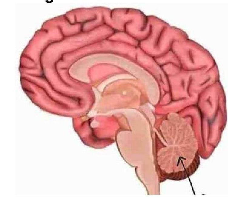
Identify the marked structure in the image.
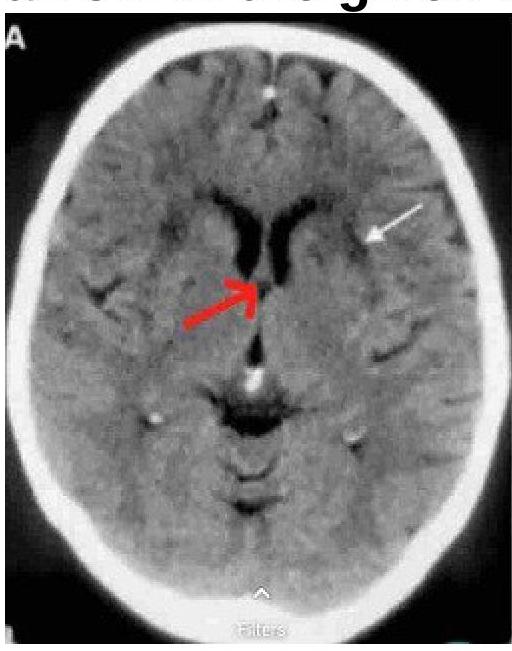
Identify the structure marked by a red arrow in the image.
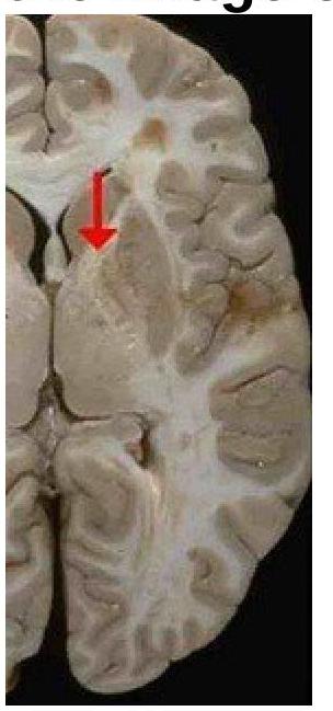
Identify the type of the fibre marked in the image of the internal capsule.
A 65-year-old lady presents with a vascular injury to the inferior frontal gyrus. Which functional area would be most affected?
Where is the auditory cortex primarily located in the brain?
Which Brodmann's area is primarily associated with motor speech?
Practice by Chapter
Cerebral Hemispheres
Practice Questions
Diencephalon
Practice Questions
Brainstem
Practice Questions
Cerebellum
Practice Questions
Basal Ganglia
Practice Questions
Limbic System
Practice Questions
Ventricular System and CSF
Practice Questions
Blood Supply of the Brain
Practice Questions
Cranial Nerves and Nuclei
Practice Questions
Functional Systems and Pathways
Practice Questions
Applied Neuroanatomy
Practice Questions
Neuroimaging Correlations
Practice Questions
Want unlimited practice?
Get full access to all questions, explanations, and performance tracking.
Scan to download app