Which of the following appears the same on both T1 and T2 weighted MRI sequences?
Which of the following investigations work on the same principle?
What is the typical MRI finding in multiple sclerosis (MS)?
Which imaging modality is most sensitive for detecting early ischemic stroke?
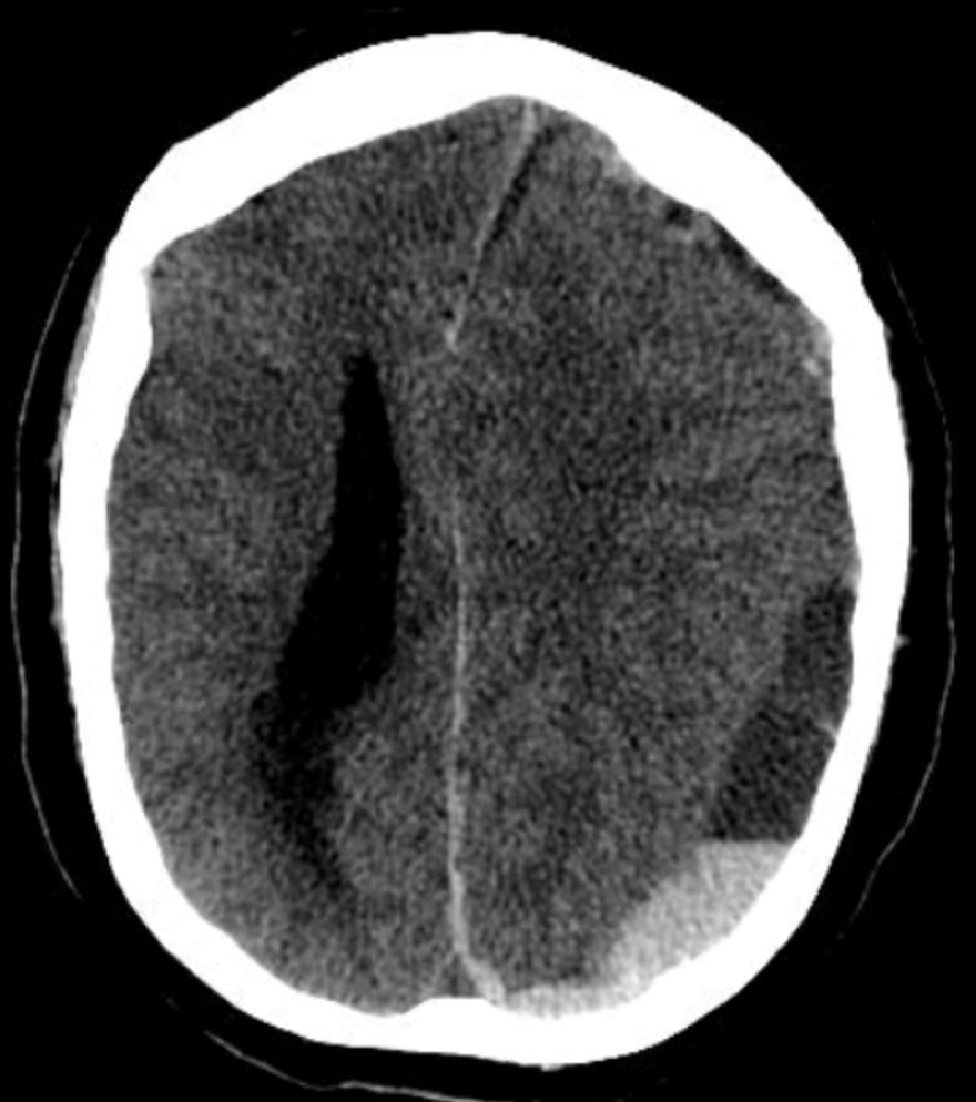
A man presents to the emergency department with a head injury following a vehicular accident. What is the investigation of choice?
A woman presenting with abrupt onset of "the worst headache of her life" Which is the best investigation?
A polytrauma patient's CT brain shows a crescent-shaped extra-axial collection with a concave inner margin. What is the most likely diagnosis?

A 40-year-old male presents with a history of headaches, fever, and new-onset seizures. An MRI of the brain is performed, revealing a ring-enhancing lesion with central restricted diffusion on diffusion-weighted imaging (DWI). What is the most likely diagnosis?
Investigation of choice for acute intracerebral hemorrhage is -
What are the typical contents of a meningocele sac?
Explanation: ***Fat*** - On both T1 and T2 weighted MRI sequences, fat appears **bright** (high signal intensity). - This consistent bright signal makes fat a useful internal reference point for signal interpretation. *Gall bladder* - The gall bladder is filled with **bile**, which appears bright on T2-weighted images due to its high water content, but can be variable on T1. - Bile does not maintain consistently the **same signal intensity** as fat on both sequences. *Kidney* - The renal parenchyma typically has **intermediate signal intensity** on both T1 and T2, but its signal characteristics are different from the consistently bright signal of fat. - The signal can vary depending on the specific sequence parameters and hydration status, unlike fat. *CSF* - **Cerebrospinal fluid (CSF)** appears dark (low signal) on T1-weighted images and bright (high signal) on T2-weighted images due to its high water content. - This distinct signal intensity difference between T1 and T2 is contrary to the shared bright appearance of fat on both sequences.
Explanation: ***CT and X-ray*** - Both **Computed Tomography (CT)** and **X-ray** imaging utilize **ionizing radiation** to generate images of the body's internal structures. - They work by passing X-ray beams through the patient, with different tissues absorbing the radiation to varying degrees, which is then detected to create an image. *MRI and PET Scan* - **Magnetic Resonance Imaging (MRI)** uses **strong magnetic fields and radio waves** to create detailed images of soft tissues, based on water content. - **Positron Emission Tomography (PET) scans** use **radioactive tracers** to visualize metabolic activity and blood flow, detecting gamma rays emitted from the patient. *CT and MRI* - **CT scans** use **ionizing radiation** (X-rays) to produce cross-sectional images. - **MRI scans** use **magnetic fields and radio waves** and do not involve ionizing radiation. *USG and HIDA Scan* - **Ultrasound (USG)** uses **high-frequency sound waves** to create real-time images of organs and structures. - **Hepatobiliary Iminodiacetic Acid (HIDA) scans** are a type of nuclear medicine study that uses a **radioactive tracer** to evaluate liver and gallbladder function.
Explanation: ***Periventricular lesions*** - **Demyelinating plaques** in a periventricular distribution are a hallmark MRI finding in **multiple sclerosis**, often appearing as **Dawson's fingers**. - These lesions reflect areas of **demyelination** and inflammation characteristic of the disease. *Basal ganglia calcification* - This finding is more commonly associated with conditions like **Fahr's disease**, **hypoparathyroidism**, or certain infections, not primary to MS. - While calcifications can occur in rare cases of MS, they are not a typical or diagnostic feature. *Diffuse cortical atrophy* - **Cortical atrophy** can be present in later stages of MS, but it is a **non-specific** finding not unique to MS and not a primary diagnostic marker. - It is more commonly seen in neurodegenerative diseases like **Alzheimer's disease** or in the elderly, and less characteristic of early MS. *Subdural hematoma* - A **subdural hematoma** is a collection of blood between the dura mater and arachnoid mater, usually due to trauma. - This is an **acute neurological emergency** and entirely unrelated to the pathology and typical MRI features of multiple sclerosis.
Explanation: ***MRI with DWI*** - **Diffusion-weighted imaging (DWI)** within an MRI scan is highly sensitive in detecting **cytotoxic edema** within minutes of **ischemic stroke** onset. This makes it crucial for early diagnosis and treatment decisions. - DWI can identify areas of restricted water diffusion, which is a hallmark of acute cellular injury due to **ischemia**, even before changes are visible on conventional T1 or T2-weighted MRI sequences. *CT* - While frequently used in acute stroke settings, **non-contrast CT** is primarily used to **rule out hemorrhagic stroke** and may only show subtle or no signs of acute ischemia in the first few hours. - Early ischemic changes on CT, often referred to as the **"ischemic penumbra"**, may appear hours after stroke onset, making it less sensitive for very early detection compared to DWI. *Ultrasound* - **Transcranial Doppler (TCD) ultrasound** can evaluate blood flow velocities in intracranial arteries and detect stenoses or occlusions but is not a primary imaging modality for directly visualizing brain parenchymal ischemia. - Cervical ultrasound (e.g., **carotid duplex**) assesses extracranial vessels but cannot directly detect **ischemic changes** within the brain tissue itself. *PET scan* - **PET (Positron Emission Tomography)** can assess brain metabolism and blood flow but is typically not the preferred or most sensitive modality for **early detection of acute ischemic stroke** due to its complexity, cost, and limited availability in emergency settings. - PET is more commonly used in research or for assessing chronic conditions and **metabolic abnormalities**, rather than acute stroke diagnosis.
Explanation: ***NCCT*** - **Non-contrast Computed Tomography (NCCT)** of the head is the **investigation of choice** for acute head trauma due to its rapid acquisition, wide availability, and excellent sensitivity for detecting acute hemorrhage, fractures, and mass effects. - It rapidly identifies life-threatening conditions such as **epidural, subdural, and intracerebral hemorrhages**, which require immediate intervention. *MRI* - **MRI** is superior for detecting subtle brain tissue injuries, diffuse axonal injury, and non-hemorrhagic lesions but is generally **not the first-line investigation** in acute trauma due to longer scan times, limited availability in the emergency setting, and inability to detect acute hemorrhage as clearly as CT. - Its use is typically reserved for follow-up studies or when CT findings are inconclusive or specific soft tissue detail is required. *CECT* - **Contrast-enhanced CT (CECT)** of the head is reserved for specific indications like evaluating vascular lesions (e.g., aneurysms, arteriovenous malformations) or tumors, which are generally **not the primary concern** in the initial assessment of acute head trauma. - Administering contrast agents can delay imaging, may pose risks to patients with renal impairment or allergies, and does not significantly improve the detection of acute traumatic hemorrhage compared to NCCT. *X-ray* - **X-rays** of the skull are useful for detecting **skull fractures**, but they provide **limited information** regarding intracranial injuries or soft tissue damage, which are critical in head trauma. - They have largely been superseded by CT scans, which offer a more comprehensive view of both bony structures and intracranial contents.
Explanation: ***NCCT of the head*** - A **non-contrast CT scan of the head** is the immediate and most appropriate first imaging study for a suspected **subarachnoid hemorrhage (SAH)**, often presenting as the "worst headache of her life" [1]. - It can rapidly detect blood in the **subarachnoid space** with high sensitivity, particularly within the first 6-12 hours of symptom onset [1], [2]. *Vessel carotid Doppler* - **Carotid Doppler ultrasound** is primarily used to assess **carotid artery stenosis** or dissection, which would not be the initial investigation for a sudden severe headache [2]. - It does not visualize intracranial blood or vascular abnormalities within the brain parenchyma or subarachnspace. *No imaging* - Given the severe, abrupt onset "worst headache of her life," **subarachnoid hemorrhage (SAH)** is a critical differential, making no imaging an inappropriate and potentially dangerous choice. - Delaying imaging could lead to severe neurological consequences if SAH is missed. *MRI* - While **MRI** can detect SAH, it is generally less accessible, takes more time to perform, and is less suitable for the initial rapid assessment of **acute SAH** compared to NCCT [3]. - **MRI** is often used for follow-up evaluation or when CT findings are equivocal, but not as the first-line emergency investigation.
Explanation: ***SDH*** - The image shows a **crescent-shaped collection** of hemorrhage with a concave inner margin, consistent with a **subdural hematoma** (SDH). - SDHs result from the tearing of **bridging veins** and typically conform to the brain's surface, crossing suture lines but not limited by bony sutures. *EDH* - An **epidural hematoma (EDH)** characteristically appears as a **lenticular** or **biconvex** shape on CT, not crescent-shaped. - EDHs are typically caused by arterial bleeding, often from the **middle meningeal artery**, and are limited by cranial sutures. *Contusion* - A **contusion** is brain tissue bruising that appears as **heterogeneous areas** of hemorrhage and edema within the brain parenchyma itself. - It would not manifest as a distinct extra-axial collection with a smooth, concave margin. *Diffuse axonal injury* - **Diffuse axonal injury (DAI)** involves widespread microscopic damage to axons, often at the gray-white matter junction. - It may appear as *punctate hemorrhages* or **small lesions** at these junctions on CT, but often the CT can be normal, and it would not present as a large extra-axial collection.
Explanation: ***Brain abscess*** - A **ring-enhancing lesion** with **central restricted diffusion** on DWI is highly characteristic of a brain abscess, due to the presence of pus containing densely packed inflammatory cells and bacteria with high viscosity. - The clinical presentation of **headaches, fever**, and **new-onset seizures** is consistent with an infectious process and increased intracranial pressure. - This combination of imaging and clinical features is pathognomonic for pyogenic brain abscess. *Glioblastoma multiforme* - While GBM can present with **ring-enhancing lesions** and seizures, it typically exhibits **facilitated diffusion** (high ADC values) on DWI due to necrotic tumor core, not restricted diffusion. - GBM is a highly infiltrative tumor with extensive **vasogenic edema**. - Fever is uncommon in GBM unless there is secondary infection. *Metastatic brain tumor* - Metastatic lesions can be **ring-enhancing** and cause seizures, but **restricted diffusion** is not typical unless there is acute hemorrhage or superimposed infection. - The presence of **fever** points away from uncomplicated metastasis. - Multiple lesions at the gray-white matter junction are more typical of metastases. *Toxoplasmosis* - Toxoplasmosis in **immunocompromised individuals** (HIV/AIDS with CD4 <100) causes **multiple ring-enhancing lesions** with predilection for basal ganglia. - Restricted diffusion is **not consistently seen** with toxoplasmosis, unlike pyogenic abscesses. - The specific DWI finding of central restricted diffusion makes brain abscess the most definitive diagnosis.
Explanation: ***NCCT*** - **Non-contrast Computed Tomography (NCCT)** is the investigation of choice for acute intracerebral hemorrhage because it can **rapidly and reliably detect acute blood** within the brain parenchyma. - Acute hemorrhage appears as a **hyperdense (bright) lesion** on NCCT, allowing for quick diagnosis and management vital in emergency settings. *MRI* - While MRI can detect hemorrhage, its sensitivity for **acute hemorrhage** can be variable, and it is **less readily available** and takes longer to perform than NCCT. - MRI is generally preferred for subacute or chronic hemorrhage, or to investigate the **underlying cause** of the bleed (e.g., tumor, vascular malformation). *PET scan* - **Positron Emission Tomography (PET) scan** primarily measures **metabolic activity** and blood flow within the brain. - It is **not suitable for detecting acute bleeding** and is typically used for diagnosing conditions like tumors, epilepsy, or neurodegenerative diseases. *None of the options* - This option is incorrect because **NCCT** is indeed the gold standard for diagnosing acute intracerebral hemorrhage.
Explanation: ***Meninges and CSF*** - A meningocele is a neural tube defect characterized by herniation of the **meninges (all three layers: dura mater, arachnoid mater, and pia mater) and cerebrospinal fluid (CSF)** through a bony defect in the skull or vertebral column. - The sac contains meninges and CSF but **does NOT contain neural tissue** (spinal cord or nerve roots), which distinguishes it from myelomeningocele. - This is typically covered by skin or a thin membrane. *Dura mater* - While the dura mater is present as the outermost layer forming part of the sac wall, it is only **one component** of the meninges. - The complete answer must include all three meningeal layers (dura, arachnoid, pia) **plus CSF**, not just the dura alone. - Stating only "dura mater" is incomplete and does not accurately describe the typical contents of a meningocele. *Spinal cord* - The presence of **spinal cord tissue** within the herniated sac indicates a more severe defect called **myelomeningocele** (or meningomyelocele). - A simple meningocele by definition does **not** contain neural tissue. *Cauda equina* - The **cauda equina** consists of spinal nerve roots below the level of L1-L2. - Its presence within the herniated sac would indicate a **myelomeningocele**, not a meningocele. - Meningocele contains only meninges and CSF, with no neural elements.
Get full access to all questions, explanations, and performance tracking.
Start For Free