Q11
The following statements are correct about burst abdomen (abdominal dehiscence) except
Q12
A patient presents to the emergency department with pain and distension of abdomen and absolute constipation. What is the investigation of choice ?
Q13
What is the investigation of choice in a patient with blunt abdominal trauma with hematuria ?
Q14
Which one of the following is the treatment of choice in a child with inguinal hernia ?
Q15
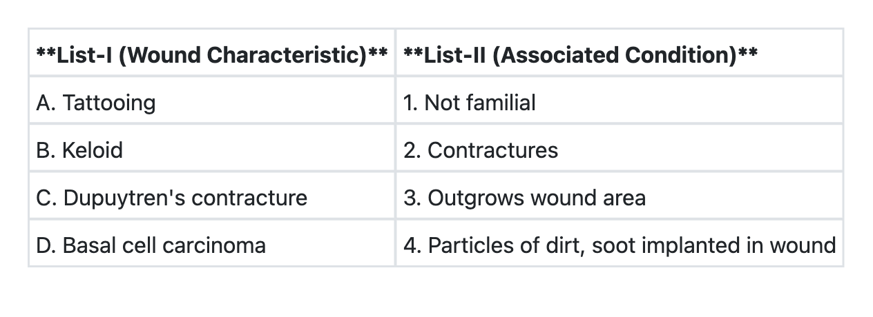
Match List-I with List-II and select the correct answer using the code given below the Lists: (Refer to the image for List-I and List-II)
Q16
Which of the following is the best indicator of prognosis of soft tissue sarcoma?
Q17
Lympho-venous anastomosis is done for
Q18
Which one of the following investigations is considered to be "Gold standard" technique for diagnosis of arterial occlusive disease ?
Q19
Which one of the following statements is not correct regarding thoracic outlet syndrome?