NEET-PG 2019 — Pathology
19 Previous Year Questions with Answers & Explanations
Fish mouth stenosis in rheumatic heart disease is due to which of the following mechanisms?
Fish mouth appearance of valve in RHD is due to-
Which marker is commonly associated with positivity in follicular lymphoma?
What are Gitter cells?
Which of the following genes primarily promotes apoptosis (programmed cell death)?
Which statement is incorrect about the pathology of the bone tumor?
Which one of the following is an autosomal recessive disorder?
A blood grouping test shows clumping with Anti-A serum, clumping with Anti-B serum, and no clumping in the control. What blood group does this indicate?
Characteristic feature of hypertrophic obstructive cardiomyopathy is:
Which chamber of the heart is enlarged first in a patient with McCallum patch?
NEET-PG 2019 - Pathology NEET-PG Practice Questions and MCQs
Question 1: Fish mouth stenosis in rheumatic heart disease is due to which of the following mechanisms?
- A. Calcification and fibrosis bridging across valvular commissures (Correct Answer)
- B. Fibrinoid necrosis
- C. Acute inflammation leading to valvular damage
- D. Myxomatous degeneration of the valve, which can occur in rheumatic heart disease
Explanation: ***Calcification and fibrosis bridging across valvular commissures*** - In rheumatic heart disease, **calcification and fibrosis** occur as a result of chronic inflammation, leading to **stenosis** of the mitral valve, often described as "fish mouth" appearance [1]. - This mechanism is due to **post-inflammatory scarring** that restricts opening and closing of the valve, characteristic of chronic rheumatic changes [1][2]. *Myxomatous degeneration of the valve* - Myxomatous degeneration primarily affects the **mitral valve** and may cause **prolapse**, but it does not lead to **stenosis**. - This process involves **thinning and elongation** of the valve leaflets, contrasting with the fibrotic changes seen in rheumatic heart disease. *Acute inflammation leading to valvular damage* - Acute inflammation due to **rheumatic fever** typically causes **valvulitis**, not chronic stenosis, which is a late consequence of chronic damage. - This mechanism leads to **valvular regurgitation** rather than stenosis, hence it's not associated with "fish mouth" stenosis. *Fibrinoid necrosis* - Fibrinoid necrosis is seen in **acute rheumatic fever** but does not directly cause **valvular stenosis**; it represents an acute inflammatory response. - This is more related to **immune complex deposition** rather than the chronic fibrotic changes leading to fish mouth morphology in rheumatic heart disease. **References:** [1] Kumar V, Abbas AK, et al.. Robbins and Cotran Pathologic Basis of Disease. 9th ed. The Heart, pp. 566-567. [2] Cross SS. Underwood's Pathology: A Clinical Approach. 6th ed. Common Clinical Problems From Cardiovascular Disease, pp. 293-294.
Question 2: Fish mouth appearance of valve in RHD is due to-
- A. Rupture of valve
- B. Calcification & fibrosis (Correct Answer)
- C. Hypertrophy of ventricular wall
- D. None of the options
Explanation: ***Calcification & fibrosis*** - The **fish mouth appearance** of the valve in rheumatic heart disease (RHD) is primarily due to **calcification and fibrosis** of the mitral valve [1]. - This results in **narrowing of the valve orifice**, which mimics the shape of a fish mouth during diastole [1]. *Rupture of valve* - Rupture of the valve typically leads to **acute severe valvular insufficiency** and does not explain the **gradual narrowing** characteristic of the fish mouth appearance. - It would generally be associated with **acute symptoms** rather than the chronic changes seen in RHD. *None of the above* - This option is incorrect as the fish mouth appearance is well-defined by **calcification and fibrosis**, making it a specific feature of RHD. - It also disregards the specific etiology associated with the valvular deformity in RHD. *Hypertrophy of ventricular wall* - While hypertrophy of the ventricular wall can occur in RHD due to increased workload, it does not directly lead to the **valvular deformity** known as fish mouth appearance. - This hypertrophy affects the **myocardium**, not the structure of the valves themselves which are primarily affected by fibrosis and calcification. **References:** [1] Kumar V, Abbas AK, et al.. Robbins and Cotran Pathologic Basis of Disease. 9th ed. The Heart, pp. 566-567.
Question 3: Which marker is commonly associated with positivity in follicular lymphoma?
- A. Bcl-1
- B. Bcl-6
- C. Bcl-2 (Correct Answer)
- D. None of the options
Explanation: ***Bcl-2*** - **Follicular lymphoma** is characterized by the overexpression of the **Bcl-2 protein**, which inhibits apoptosis, leading to the survival of malignant B cells [1][3]. - The **Bcl-2 gene** is often involved in the **t(14;18)** chromosomal translocation, which is a hallmark of this lymphoma [1][3][4]. *Bcl-6* - Although **Bcl-6** can be expressed in some lymphomas, it is primarily associated with **diffuse large B-cell lymphoma**, not follicular lymphoma. - **Bcl-6** is involved in **germinal center formation** and its positivity does not indicate follicular lymphoma specifically. *Bcl-1* - **Bcl-1** (also known as **CCND1**) is primarily associated with **mantle cell lymphoma** and is not a characteristic marker for follicular lymphoma. - It is linked to the **t(11;14)** translocation, which is distinct from the genetic alterations seen in follicular lymphoma. *None of the above* - This option is incorrect as **Bcl-2 positivity** is definitive for follicular lymphoma [2]. - The presence of other markers like **Bcl-6** or **Bcl-1** does not negate the expression of Bcl-2 in this lymphoma type. **References:** [1] Kumar V, Abbas AK, et al.. Robbins and Cotran Pathologic Basis of Disease. 9th ed. Diseases of White Blood Cells, Lymph Nodes, Spleen, and Thymus, pp. 602-604. [2] Kumar V, Abbas AK, et al.. Robbins and Cotran Pathologic Basis of Disease. 9th ed. Diseases of White Blood Cells, Lymph Nodes, Spleen, and Thymus, p. 604. [3] Cross SS. Underwood's Pathology: A Clinical Approach. 6th ed. Common Clinical Problems From Diseases Of The Urinary And Male Genital Tracts, pp. 561-562. [4] Kumar V, Abbas AK, et al.. Robbins and Cotran Pathologic Basis of Disease. 9th ed. Neoplasia, pp. 310-311.
Question 4: What are Gitter cells?
- A. Microglia
- B. Modified macrophages in the CNS (Correct Answer)
- C. Astrocytic cells
- D. Oligodendrocytic cells
Explanation: ***Modified macrophages in CNS*** - Gitter cells are **modified macrophages** that have phagocytized lipid and other debris in the central nervous system (CNS), particularly in response to injury or disease [1][2]. - They play a crucial role in **cleaning up cellular debris** and are involved in the inflammatory response within the CNS [2]. *Macroglia* - Macroglia refers to **supportive cells** in the CNS, including astrocytes and oligodendrocytes, rather than being specifically modified macrophages. - It does not specifically describe the **phagocytic role** characteristic of Gitter cells. *Oligodendrocytes* - Oligodendrocytes primarily function to **myelinate axons** in the CNS and do not possess the same phagocytic capabilities as Gitter cells. - They are involved in **insulation** of neuronal axons rather than debris clearance. *Astrocytes* - Astrocytes are the principal **supportive glial cells** in the CNS and do not exhibit the characteristics of Gitter cells. - Their functions include **maintaining blood-brain barrier**, regulating blood flow, and supporting neuronal metabolism, not phagocytosis. **References:** [1] Kumar V, Abbas AK, et al.. Robbins and Cotran Pathologic Basis of Disease. 9th ed. Peripheral Nerves and Skeletal Muscles, pp. 1255-1256. [2] Cross SS. Underwood's Pathology: A Clinical Approach. 6th ed. Common Clinical Manifestations Of Central And Peripheral Nervous System Disease, pp. 697-698.
Question 5: Which of the following genes primarily promotes apoptosis (programmed cell death)?
- A. Bax (Correct Answer)
- B. Bclx
- C. Mcl
- D. Bcl2
Explanation: ***Bax*** - **Bax** is a **pro-apoptotic gene** that actively promotes programmed cell death by forming channels in the mitochondrial outer membrane, leading to cytochrome c release and activation of caspases [1]. - It is a key member of the Bcl-2 family and plays a critical role in the **intrinsic apoptotic pathway** [2]. - Unlike the other options, Bax **promotes** rather than inhibits apoptosis [1]. *Bclx* - **Bcl-xL** is an **anti-apoptotic** gene that **inhibits** cell death by preventing the activation of pro-apoptotic proteins like Bax [1]. - It maintains mitochondrial membrane integrity, thereby **blocking** apoptosis [1]. *Mcl* - **Mcl-1** (Myeloid cell leukemia sequence 1) is an **anti-apoptotic** gene belonging to the Bcl-2 family [1]. - Its primary role is to **inhibit** apoptosis and promote cell survival by sequestering pro-apoptotic proteins [1]. *Bcl2* - **Bcl-2** (B-cell lymphoma 2) is the prototype **anti-apoptotic** gene that **prevents** programmed cell death [3]. - It functions by binding to and inhibiting pro-apoptotic members of the Bcl-2 family, thus maintaining cell viability and **opposing** apoptosis [3]. **References:** [1] Kumar V, Abbas AK, et al.. Robbins and Cotran Pathologic Basis of Disease. 9th ed. Neoplasia, p. 310. [2] Kumar V, Abbas AK, et al.. Robbins and Cotran Pathologic Basis of Disease. 9th ed. Cellular Responses to Stress and Toxic Insults: Adaptation, Injury, and Death, pp. 64-67. [3] Kumar V, Abbas AK, et al.. Robbins and Cotran Pathologic Basis of Disease. 9th ed. Neoplasia, pp. 310-311.
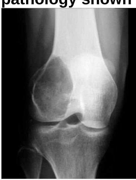
Question 6: Which statement is incorrect about the pathology of the bone tumor?
- A. Tumor has distinct margin
- B. Tumor arises from epiphyseal to metaphyseal region
- C. Eccentric lesion
- D. Chemotherapy is the treatment of choice for all bone tumors. (Correct Answer)
Explanation: ***Tumor has distinct margin*** - A **distinct margin** often indicates a benign tumor, while malignant tumors typically show **infiltrative margins**. - In bone tumors, particularly malignant ones, the lack of clear demarcation is a key pathological feature. *Chemotherapy is the treatment of choice* - While chemotherapy may be used for certain **malignant bone tumors**, it is not the first-line treatment for most bone tumors [1]. - The primary treatment is often **surgical excision**, especially for localized lesions [1]. *Tumor arise from epiphyseal to metaphyseal region* - While some tumors can originate in these areas, many actually arise from the **diaphyseal** region in bone tumors like osteosarcoma. - This option misrepresents the common locations where various tumors develop, as osteochondromas tend to develop near the epiphyses of limb bones [2]. *Eccentric lesion* - Many bone tumors do indeed present as **eccentric lesions**, especially benign ones like **osteochondromas**. - However, this feature does not apply universally, as some malignant tumors can also be **central or infiltrative** in nature. **References:** [1] Cross SS. Underwood's Pathology: A Clinical Approach. 6th ed. Common Clinical Problems From Osteoarticular And Connective Tissue Disease, pp. 673-674. [2] Cross SS. Underwood's Pathology: A Clinical Approach. 6th ed. Common Clinical Problems From Osteoarticular And Connective Tissue Disease, pp. 672-673.
Question 7: Which one of the following is an autosomal recessive disorder?
- A. Albinism (Correct Answer)
- B. Marfan’s syndrome
- C. Neurofibromatosis-1
- D. Huntington's disease
Explanation: ***Albinism*** - **Albinism** is an **autosomal recessive disorder** characterized by a partial or complete lack of melanin pigment in the skin, hair, and eyes [1], [2]. - This condition is inherited when an individual receives **two copies of the defective gene**, one from each parent [1]. *Huntington's disease* - **Huntington's disease** is an **autosomal dominant disorder**, meaning only one copy of the mutated gene is sufficient to cause the disease. - It is characterized by progressive neurodegeneration, leading to uncontrolled movements, cognitive decline, and psychiatric problems. *Marfan's syndrome* - **Marfan's syndrome** is an **autosomal dominant disorder** affecting connective tissue, primarily impacting the skeletal, ocular, and cardiovascular systems. - It results from a mutation in the **FBN1 gene** which encodes for fibrillin-1, a component of elastic fibers. *Neurofibromatosis-1* - **Neurofibromatosis type 1 (NF1)** is an **autosomal dominant disorder** caused by a mutation in the NF1 gene, leading to the growth of tumors along nerves. - Clinical features include **café-au-lait spots**, neurofibromas, and Lisch nodules. **References:** [1] Kumar V, Abbas AK, et al.. Robbins and Cotran Pathologic Basis of Disease. 9th ed. Genetic Disorders, pp. 150-151. [2] Cross SS. Underwood's Pathology: A Clinical Approach. 6th ed. (Basic Pathology) introduces the student to key general principles of pathology, both as a medical science and as a clinical activity with a vital role in patient care. Part 2 (Disease Mechanisms) provides fundamental knowledge about the cellular and molecular processes involved in diseases, providing the rationale for their treatment. Part 3 (Systematic Pathology) deals in detail with specific diseases, with emphasis on the clinically important aspects., pp. 119-120.
Question 8: A blood grouping test shows clumping with Anti-A serum, clumping with Anti-B serum, and no clumping in the control. What blood group does this indicate?
- A. A
- B. B
- C. O
- D. AB (Correct Answer)
Explanation: ***AB*** - The results show **clumping with both Anti-A and Anti-B serum**, indicating the presence of both A and B antigens on the red blood cells. - The absence of clumping in the control confirms that the **agglutination with Anti-A and Anti-B is due to specific antigen-antibody reactions**, not nonspecific agglutination. - Blood group AB individuals have both A and B antigens on their RBCs and no anti-A or anti-B antibodies in their serum. *A* - Blood group A would show **clumping with Anti-A serum only** and no clumping with Anti-B serum. - This is incorrect because the sample shows clumping with both antisera. *B* - Blood group B would show **clumping with Anti-B serum only** and no clumping with Anti-A serum. - This is incorrect because the sample shows clumping with both antisera. *O* - Blood group O would show **no clumping with either Anti-A or Anti-B serum**, as it lacks both A and B antigens. - This is incorrect because the sample clearly shows clumping with both Anti-A and Anti-B sera.
Question 9: Characteristic feature of hypertrophic obstructive cardiomyopathy is:
- A. Increased size of ventricle
- B. Asymmetric septal hypertrophy (Correct Answer)
- C. Normal myofiber arrangement
- D. Increased size of atria
Explanation: ***Asymmetric septal hypertrophy*** - This is the hallmark feature of **hypertrophic obstructive cardiomyopathy (HOCM)**, where the **interventricular septum** thickens disproportionately more than the free wall of the left ventricle [1], [2]. - This septal thickening can lead to **left ventricular outflow tract obstruction**, particularly during systole, obstructing blood flow out of the heart [1]. *Increased size of ventricle* - While the left ventricle may appear enlarged in some dimensions due to hypertrophy, the primary characteristic is specifically **asymmetric thickening of the septum**, not a generalized increase in ventricular size [2]. - In other forms of cardiomyopathy, such as dilated cardiomyopathy, a global increase in ventricular size is observed, which is distinct from HOCM. *Normal myofiber arrangement* - A characteristic microscopic feature of HOCM is **myocardial disarray**, where cardiac muscle fibers are abnormally arranged instead of their usual parallel alignment [1]. - This disorganized arrangement contributes to the impaired function and electrical instability seen in HOCM. *Increased size of atria* - While **left atrial enlargement** can be a secondary finding in HOCM due to increased left ventricular end-diastolic pressure and impaired diastolic filling, it is not the primary or characteristic feature defining the condition [1]. - The fundamental pathology of HOCM lies in the specific hypertrophy of the ventricular myocardium, particularly the septum. **References:** [1] Kumar V, Abbas AK, et al.. Robbins and Cotran Pathologic Basis of Disease. 9th ed. The Heart, pp. 577-578. [2] Cross SS. Underwood's Pathology: A Clinical Approach. 6th ed. Common Clinical Problems From Cardiovascular Disease, pp. 303-304.
Question 10: Which chamber of the heart is enlarged first in a patient with McCallum patch?
- A. Left atrium (Correct Answer)
- B. Left ventricle
- C. Right atrium
- D. Right ventricle
Explanation: ***Left atrium*** - A **McCallum patch** is a thickened, often irregular endocardial lesion found in the **left atrium**. - It results from the jet lesion of **mitral regurgitation**, indicating the left atrium has been subjected to increased volume and pressure leading to enlargement [1]. *Left ventricle* - While **mitral regurgitation** can eventually lead to **left ventricular enlargement** (due to volume overload), the primary chamber affected by the regurgitant jet causing the McCallum patch is the left atrium. - Left ventricular enlargement is a later consequence, not the first chamber to show this specific lesion. *Right atrium* - The **right atrium** is affected by conditions like **tricuspid regurgitation** or **pulmonary hypertension**, which are unrelated to mitral valve disease or McCallum patches. - It handles systemic venous return, separate from the left-sided circulation involved in mitral pathology. *Right ventricle* - The **right ventricle** is primarily impacted by conditions affecting the **pulmonary circulation** or **tricuspid valve**. - It fills from the right atrium and pumps blood to the lungs, making it unlikely to be the first chamber enlarged in the context of a McCallum patch from mitral regurgitation. **References:** [1] Kumar V, Abbas AK, et al.. Robbins and Cotran Pathologic Basis of Disease. 9th ed. Diseases of Infancy and Childhood, pp. 533-534.