All (314)Anatomy (1)Anatomy (26)Anesthesiology (9)Biochemistry (26)Community Medicine (10)Dermatology (15)ENT (7)Forensic Medicine (5)General Medicine (1)Internal Medicine (36)Microbiology (23)Obstetrics and Gynecology (13)Ophthalmology (10)Orthopaedics (6)Pathology (1)Pathology (25)Pediatrics (13)Pharmacology (37)Physiology (15)Psychiatry (2)Psychiatry (4)Radiology (11)Surgery (18)
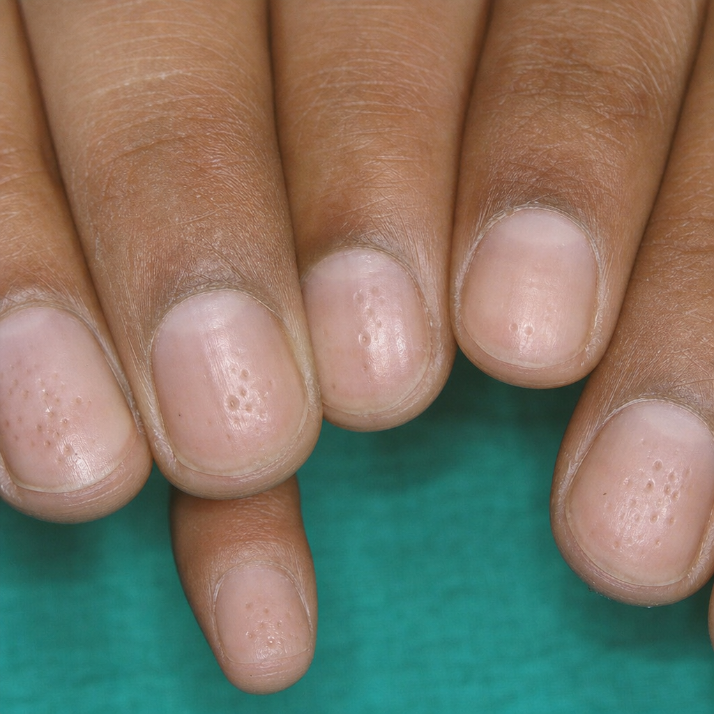
Q141
Which of the following nail findings is seen in the condition shown below?
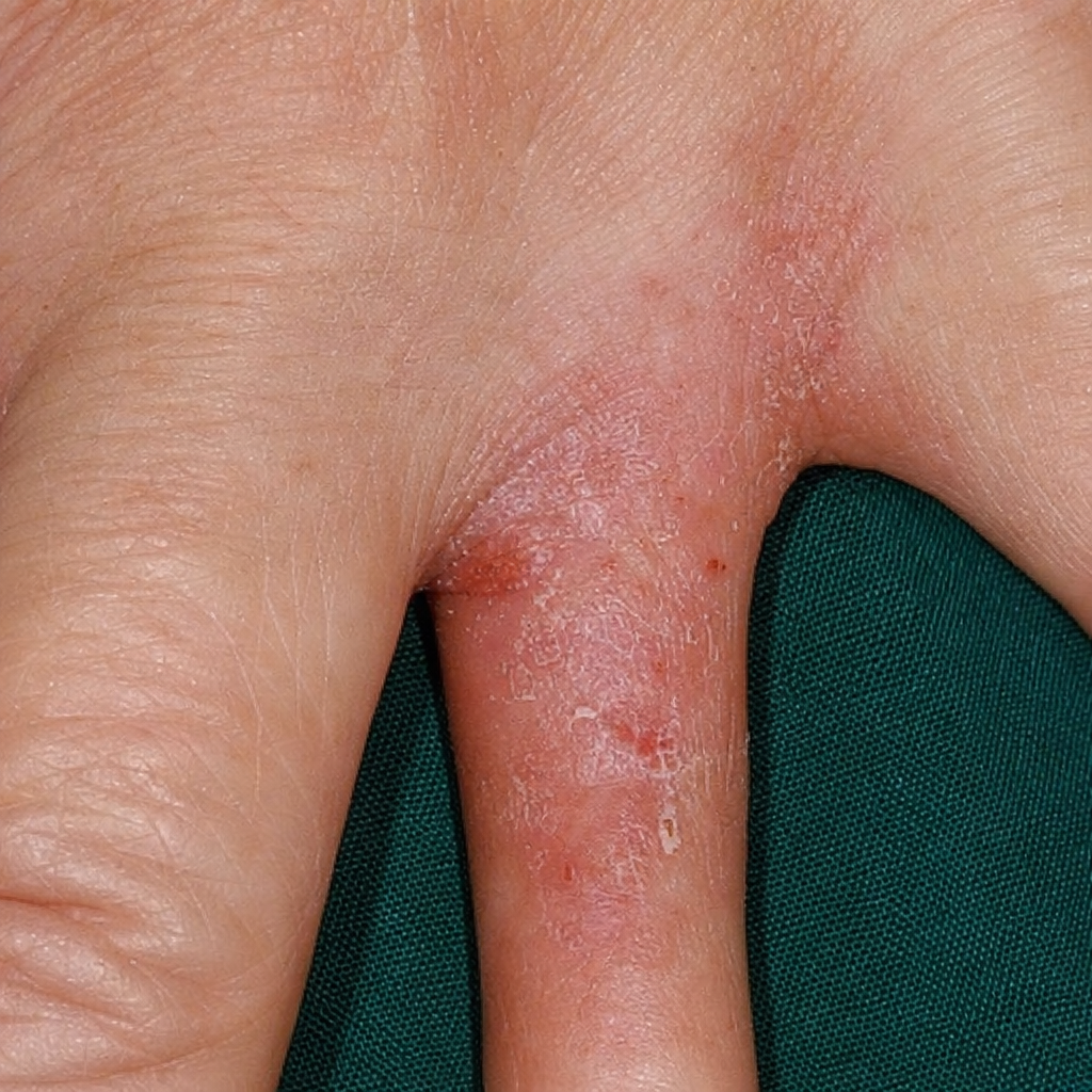
Q142
Patient presents with pruritus of the interdigital spaces of the left hand as shown in the image. Identify the condition.