NEET-PG 2019 — Orthopaedics
6 Previous Year Questions with Answers & Explanations
Which part of scaphoid fracture is most susceptible to avascular necrosis?
What should be the most likely diagnosis of this 65-year-old lady who presents with backache?
A 25-year-old male presents with localized pain in the tibia and swelling. Imaging reveals a bone abscess. Identify the condition.
Scaphoid fracture which area has maximum chances of AVN/Non-union/Malunion:-
12 years male came with swelling of lower end tibia which is surrounded by rim of reactive bone. What is most likely diagnosis?
A patient with GCT, which of the following is false?
NEET-PG 2019 - Orthopaedics NEET-PG Practice Questions and MCQs
Question 1: Which part of scaphoid fracture is most susceptible to avascular necrosis?
- A. Distal 1/3rd
- B. Middle 1/3rd
- C. Proximal 1/3rd (Correct Answer)
- D. Scaphoid Tubercle
Explanation: ***Proximal 1/3rd*** - The **scaphoid bone** has a **retrograde blood supply**, meaning blood vessels enter distally and flow towards the proximal pole. - A fracture in the **proximal 1/3rd** can disrupt the blood supply to the **proximal fragment**, making it highly susceptible to **avascular necrosis**. *Distal 1/3rd* - Fractures in the **distal 1/3rd** of the scaphoid generally have a robust blood supply due to the entry of vessels from the distal pole. - While still requiring proper management, the risk of **avascular necrosis** is significantly lower compared to proximal fractures. *Middle 1/3rd* - Fractures in the **middle 1/3rd** (waist) of the scaphoid are common and can still compromise blood flow to the proximal segment, but the risk of **avascular necrosis** is intermediate. - The more proximal the fracture within the middle third, the higher the risk of **avascular necrosis**. *Scaphoid Tubercle* - The **scaphoid tubercle** is a distal projection of the scaphoid bone. - Fractures of the **scaphoid tubercle** are extra-articular and typically have an excellent blood supply; thus, they are at very low risk for **avascular necrosis**.
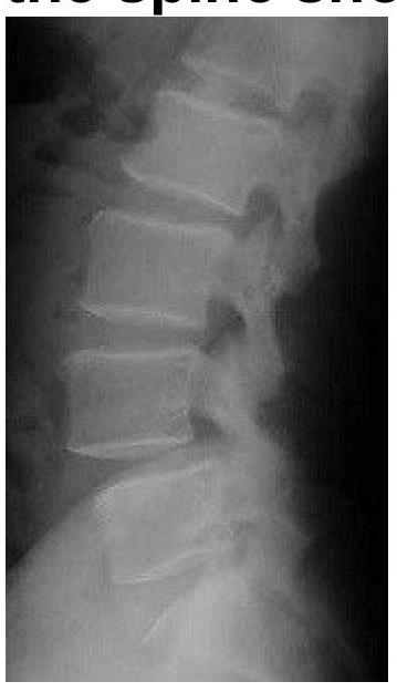
Question 2: What should be the most likely diagnosis of this 65-year-old lady who presents with backache?
- A. Osteoporotic fracture
- B. Spondylolisthesis (Correct Answer)
- C. Spondylolysis
- D. Discitis
Explanation: ***Spondylolisthesis*** - The lateral X-ray image reveals an **anterior displacement of one vertebral body over the one below it**, which is characteristic of spondylolisthesis. - In a 65-year-old lady, degenerative spondylolisthesis due to **arthritic changes and instability** is a common cause of backache. *Osteoporotic fracture* - An osteoporotic fracture would typically show a **compression deformity** or a wedge-shaped vertebral body, which is not clearly depicted here. - While osteoporosis is common in this age group, the primary finding on this image is vertebral slippage, not fracture. *Spondylolysis* - Spondylolysis is a **defect in the pars interarticularis** (a thin segment of bone connecting the superior and inferior articular facets) and is best seen on oblique views or CT. - Although spondylolysis can *lead to* spondylolisthesis, the immediate and most striking finding on this lateral view is the slippage itself. *Discitis* - Discitis, an **inflammation or infection of the intervertebral disc and adjacent vertebrae**, would typically show **loss of disc height** and **endplate irregularities or erosions**. - These features are not the predominant finding on this image, which clearly demonstrates vertebral body displacement.
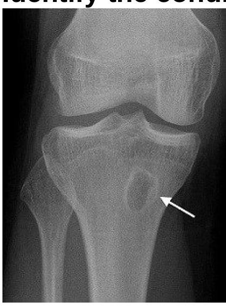
Question 3: A 25-year-old male presents with localized pain in the tibia and swelling. Imaging reveals a bone abscess. Identify the condition.
- A. Brodie abscess (Correct Answer)
- B. Osteoid osteoma
- C. Intracortical hemangioma
- D. Chondromyxoid fibroma
Explanation: ***Brodie abscess*** - A Brodie abscess is a **subacute or chronic osteomyelitis** characterized by a well-circumscribed, **radiolucent lesion** (an abscess cavity) often surrounded by a zone of **sclerosis**, representing the body's attempt to wall off the infection. - The presentation of localized pain and swelling in the tibia, with imaging revealing a bone abscess, is consistent with this condition, which is a common form of localized osteomyelitis. *Osteoid osteoma* - This is a **benign bone tumor** characterized by a small, radiolucent nidus surrounded by a large area of **sclerotic bone**. The pain from an osteoid osteoma is typically **worse at night** and dramatically relieved by NSAIDs. - While it can cause localized pain and swelling, the imaging features of a distinct abscess cavity are not characteristic of an osteoid osteoma. *Intracortical hemangioma* - An intracortical hemangioma is a **rare benign vascular lesion** within the cortex of a bone. - Imaging typically shows a **lytic lesion** with a characteristic **"honeycomb" or "sunburst" appearance**, not a well-defined abscess. *Chondromyxoid fibroma* - This is a rare, **benign cartilaginous tumor** that usually presents as an **eccentric lytic lesion** in the metaphysis of long bones, often with a scalloped border and sclerotic rim. - While it can cause localized pain and swelling, the imaging appearance of an abscess with sclerotic margins is not typical of a chondromyxoid fibroma.
Question 4: Scaphoid fracture which area has maximum chances of AVN/Non-union/Malunion:-
- A. Distal 1/3
- B. Proximal 1/3 (Correct Answer)
- C. Scaphoid Tubercle fracture
- D. Middle 1/3
Explanation: ***Proximal 1/3*** - The **proximal pole of the scaphoid** has a precarious blood supply, primarily from retrograde extraosseous vessels entering distally. A fracture in this region can compromise this supply, leading to **avascular necrosis (AVN)**. - Due to the limited blood flow to the proximal fragment, healing is often impaired, increasing the risk of **non-union** and **malunion**. *Distal 1/3* - Fractures in the **distal 1/3 (distal pole)** of the scaphoid typically have a better prognosis. - This area has a more robust blood supply, reducing the risk of AVN and promoting faster healing. *Scaphoid Tubercle fracture* - Fractures of the **scaphoid tubercle** are usually considered stable and intra-articular, with a good blood supply. - These fractures generally heal well with conservative treatment and have a very low incidence of AVN or non-union. *Middle 1/3* - Fractures in the **middle 1/3 (waist)** of the scaphoid are the most common but still pose a significant risk of non-union. - While the risk of AVN is lower than for proximal pole fractures, it is still higher than for distal fractures, due to the critical vascular supply to both fragments.
Question 5: 12 years male came with swelling of lower end tibia which is surrounded by rim of reactive bone. What is most likely diagnosis?
- A. GCT
- B. Hyper PTH
- C. Brodie's Abscess (Correct Answer)
- D. Osteomyelitis
Explanation: ***Brodie's Abscess*** - A **Brodie's abscess** is a subacute or chronic osteomyelitis characterized by a localized bone abscess, typically with a surrounding **sclerotic rim of reactive bone**. - It often occurs in the **metaphysis of long bones** (like the lower end of the tibia) in children and adolescents, presenting with localized pain and swelling. *GCT* - **Giant cell tumor (GCT)** typically occurs in **skeletally mature adults** (20-40 years old) and is a lytic lesion often found in the **epiphysis** of long bones, rarely with a distinct sclerotic rim. - GCTs are generally more aggressive and demonstrate a **soap-bubble appearance** with cortical expansion rather than a thick reactive bone rim. *Hyper PTH* - **Hyperparathyroidism** causes bone changes such as **osteopenia**, **subperiosteal bone resorption**, especially in the phalanges, and **brown tumors** (lytic lesions). - It does not typically present as a localized lesion with a **sclerotic rim of reactive bone** in a child. *Osteomyelitis* - While chronic osteomyelitis can involve local bone destruction and reactive bone formation, a **Brodie's abscess** is a specific, well-circumscribed form of **subacute osteomyelitis**. - Acute osteomyelitis presents with more diffuse systemic symptoms (fever, malaise) and less defined reactive bone in its early stages compared to the distinct **sclerotic rim** seen in a Brodie's abscess.
Question 6: A patient with GCT, which of the following is false?
- A. Defined margins
- B. Chemotherapy is the mainstay of treatment (Correct Answer)
- C. Epiphyseo-metaphyseal location
- D. Eccentric
Explanation: ***Chemotherapy is the mainstay of treatment*** - This statement is **false** because **Giant Cell Tumor of Bone (GCT)** therapy primarily involves **surgical resection**, with or without adjuvant therapies like **denosumab**. - **Chemotherapy** is generally *not* the first-line treatment for GCT, as these tumors respond poorly to it; it's usually reserved for cases of **metastatic GCT** or when other treatments fail. *Defined margins* - GCTs often present radiographically with **well-defined, non-sclerotic margins**, which indicates a lytic lesion that is often locally aggressive but typically doesn't invade widely. - While they are locally destructive, their borders are usually visible, helping distinguish them from other bone tumors. *Epiphyseo-metaphyseal location* - GCTs commonly originate in the **metaphysis** of long bones and **extend into the epiphysis** after the growth plate has closed. - This characteristic location near a joint is a classic diagnostic feature of GCT, especially in adults. *Eccentric* - GCTs typically arise **eccentrically** within the bone, meaning they originate off-center in the bone marrow cavity before expanding and thinning the cortex. - This eccentric growth pattern is a distinguishing feature, particularly in contrast to other bone tumors which might be centrally located.