NEET-PG 2019 — Ophthalmology
7 Previous Year Questions with Answers & Explanations
Esotropia is most commonly associated with:
What conditions can be diagnosed using the cover-uncover test?
Esotropia is commonly seen in which type of refractive error?
Which is the most common ocular finding in myasthenia gravis?
Which of the following statements about conjunctival lesions is NOT true?
The most common cause of proptosis in adults:-
Identify the instrument? (NEET Pattern 2019)
NEET-PG 2019 - Ophthalmology NEET-PG Practice Questions and MCQs
Question 1: Esotropia is most commonly associated with:
- A. Hyperopia (Correct Answer)
- B. Presbyopia
- C. Astigmatism
- D. Myopia
Explanation: ***Hyperopia*** - **Hyperopia** (farsightedness) requires greater accommodative effort to focus on distant and near objects, which is coupled with **convergence**. This excessive convergence can lead to **esotropia** (inward turning of the eye). - Accommodative esotropia is a common type of strabismus directly linked to uncorrected hyperopia. *Presbyopia* - **Presbyopia** is an age-related loss of the eye's ability to focus on nearby objects due to stiffening of the lens, typically occurring after age 40. - It affects accommodation but does not primarily cause esotropia; rather, it makes near work difficult, and patients may prefer to hold objects further away to see them. *Astigmatism* - **Astigmatism** is a refractive error where the eye does not focus light evenly onto the retina due to an irregularly shaped cornea or lens, leading to blurred or distorted vision at all distances. - While it can cause visual discomfort and eye strain, it is not directly associated with the development of esotropia. *Myopia* - **Myopia** (nearsightedness) is a refractive error where distant objects appear blurry because light focuses in front of the retina. - High myopia can sometimes be associated with **exotropia** (outward turning of the eye) due to divergence excess, rather than esotropia.
Question 2: What conditions can be diagnosed using the cover-uncover test?
- A. Eye alignment disorders including strabismus and heterophoria (Correct Answer)
- B. Convergent strabismus (Esotropia)
- C. Latent misalignment (Heterophoria)
- D. Strabismus (Squint)
Explanation: ***Eye alignment disorders including strabismus and heterophoria*** - The **cover-uncover test** is a clinical procedure used to detect and differentiate both **strabismus** (manifest deviation) and **heterophoria** (latent deviation) by observing eye movements when vision is occluded and then re-exposed. - This test is a fundamental tool for assessing **ocular alignment** and binocular vision, revealing if an eye deviates and how it recovers. - **This is the most comprehensive answer** as it includes both manifest and latent deviations. *Convergent strabismus (Esotropia)* - Although the cover-uncover test can diagnose **esotropia** (a type of strabismus where the eye turns inward), this option is **too specific** and does not cover all the conditions assessable by this test. - The test can diagnose **all types of strabismus** (esotropia, exotropia, hypertropia, hypotropia) and heterophoria, not just convergent strabismus. - Esotropia is characterized by the **deviating eye failing to spontaneously realign** when uncovered, as it is a constant, manifest deviation. *Latent misalignment (Heterophoria)* - While the cover-uncover test **can detect heterophoria**, this option is **incomplete** as it does not include strabismus (manifest deviation). - Heterophoria manifests when the covered eye deviates and then **refixes** when uncovered, indicating a latent deviation normally controlled by fusion. - The alternate cover test is more sensitive for detecting heterophoria, but the cover-uncover test can identify it as well. *Strabismus (Squint)* - The cover-uncover test is used to diagnose **strabismus**, but this option is **incomplete** and does not include **heterophoria**, which is also diagnosable by the test. - Strabismus is identified when the eye that was *not* covered deviates, or the covered eye does not refixate upon uncovering, indicating a manifest turn. - This option only covers manifest deviations and misses latent deviations.
Question 3: Esotropia is commonly seen in which type of refractive error?
- A. Myopia
- B. Hypermetropia (Correct Answer)
- C. Astigmatism
- D. Presbyopia
Explanation: ***Hypermetropia*** - **Esotropia**, or convergent strabismus, is commonly associated with **uncorrected hypermetropia**, especially in children. - The constant effort to **accommodate** to see clearly for hypermetropic individuals can lead to excessive convergence, causing the eye to turn inward. *Myopia* - Myopia, or **nearsightedness**, rarely causes esotropia. - In some cases, high myopia can be associated with **exotropia** (divergent strabismus) due to reduced accommodative effort. *Astigmatism* - **Astigmatism** causes blurry vision at all distances due to an irregularly shaped cornea or lens, but it is not directly linked to specific forms of strabismus like esotropia or exotropia. - While it can contribute to **amblyopia** if severe and uncorrected, it does not typically cause the eyes to turn inward. *Presbyopia* - **Presbyopia** is an age-related loss of the eye's ability to focus on nearby objects due to stiffening of the lens. - It affects accommodation but does not cause strabismus such as esotropia; it typically begins around age 40.
Question 4: Which is the most common ocular finding in myasthenia gravis?
- A. Ptosis (Correct Answer)
- B. Lagophthalmos
- C. Proptosis
- D. Enophthalmos
Explanation: ***Ptosis*** - **Ptosis**, or drooping of the eyelid, is the most common ocular manifestation of **myasthenia gravis**, affecting a large majority of patients. - It results from **weakness of the levator palpebrae superioris muscle**, which is responsible for lifting the eyelid. *Lagophthalmos* - **Lagophthalmos** is the inability to close the eyelids completely, often due to facial nerve palsy or severe proptosis. - While it can lead to exposure keratopathy, it is **not a primary or common finding** in myasthenia gravis. *Proptosis* - **Proptosis** (or exophthalmos) is the forward bulging of the eyeball, most commonly associated with **Graves' ophthalmopathy**. - It is **not a feature of myasthenia gravis**, which typically involves muscle weakness, not orbital mass effects. *Enophthalmos* - **Enophthalmos** refers to the posterior displacement of the eyeball within the orbit, often seen in conditions like **orbital fractures** or Horner's syndrome. - It is **not associated with the neuromuscular dysfunction** characteristic of myasthenia gravis.
Question 5: Which of the following statements about conjunctival lesions is NOT true?
- A. Arise from any part of conjunctiva
- B. Can cause Astigmatism
- C. Surgery is treatment of choice (Correct Answer)
- D. UV exposure is risk factor
Explanation: ***Surgery is treatment of choice*** - While surgery can be used to treat conjunctival lesions, it is not always the **treatment of choice**, especially for smaller, asymptomatic lesions like **pinguecula** which may only require observation and lubrication. - Many conjunctival lesions, such as uncomplicated **pterygium** or **pinguecula**, are managed conservatively unless they cause significant symptoms, vision impairment, or cosmetic concerns. *Arise from any part of conjunctiva* - **Conjunctival lesions** can indeed arise from various parts of the conjunctiva, including the palpebral, bulbar, and forniceal conjunctiva. - For example, **pterygium** typically arises from the bulbar conjunctiva, while **pinguecula** also originates in the bulbar conjunctiva, specifically in the interpalpebral fissure. *Can cause Astigmatism* - Larger **conjunctival lesions**, particularly a **pterygium** that encroaches onto the cornea, can induce or alter astigmatism. - The growth of the lesion can change the **curvature of the cornea**, leading to optical distortion and astigmatism. *UV exposure is risk factor* - **Ultraviolet (UV) light exposure** is a well-established risk factor for the development of many conjunctival lesions, including **pterygium** and **pinguecula**. - Chronic UV exposure leads to **elastotic degeneration** of the conjunctival collagen and is thought to play a key role in the pathogenesis of these growths.
Question 6: The most common cause of proptosis in adults:-
- A. Preseptal cellulitis
- B. Capillary hemangioma
- C. Thyroid eye disease (Correct Answer)
- D. Orbital cellulitis
Explanation: ***Thyroid eye disease*** - **Thyroid eye disease (TED)**, also known as Graves' ophthalmopathy, is the most common cause of **proptosis** in adults. - It results from an autoimmune process leading to inflammation and expansion of the **extraocular muscles** and orbital fat, which pushes the eyeball forward. *Preseptal cellulitis* - **Preseptal cellulitis** is an infection of the eyelid and periorbital tissue anterior to the orbital septum, typically presenting with **eyelid swelling** and redness. - While it causes periorbital swelling, it generally does not cause true **proptosis**, which is the anterior displacement of the eyeball itself. *Capillary hemangioma* - **Capillary hemangiomas** are benign vascular tumors and are the most common orbital tumor in **infancy and childhood**, not adults. - They typically cause proptosis in young children, often presenting as a **reddish-blue mass** that may increase in size with crying. *Orbital cellulitis* - **Orbital cellulitis** is a serious infection of the tissues within the orbit, posterior to the orbital septum, which can cause **proptosis**, pain, and ophthalmoplegia. - While it is a cause of proptosis, it is an **acute infectious process** and not the most common overall cause of proptosis in the adult population compared to thyroid eye disease.
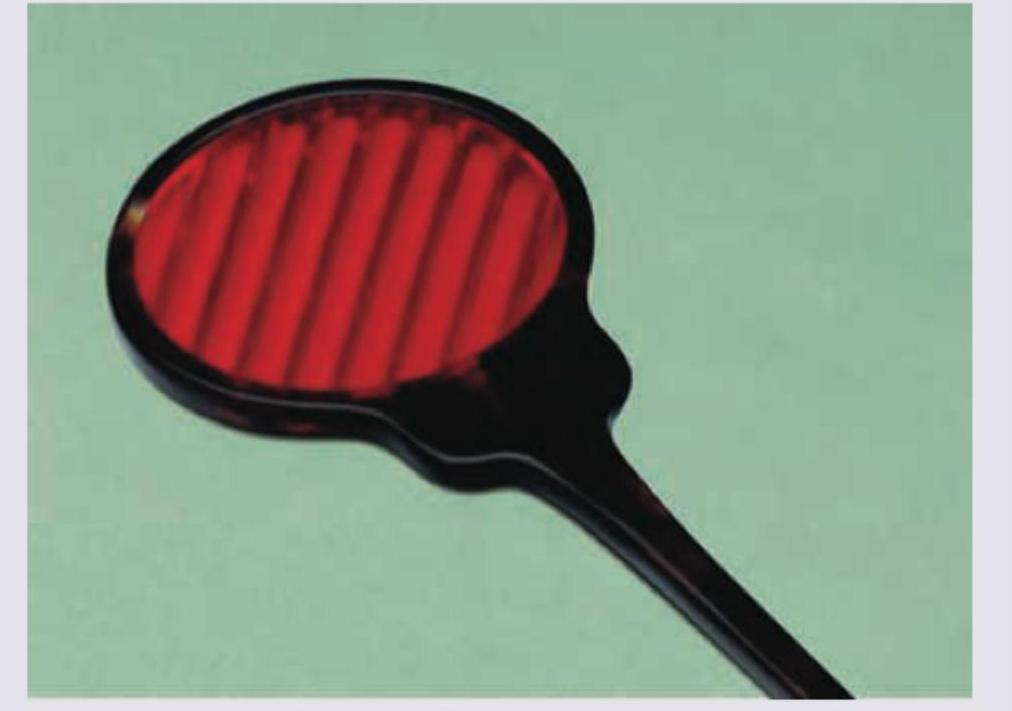
Question 7: Identify the instrument? (NEET Pattern 2019)
- A. Maddox rod (Correct Answer)
- B. Maddox wing
- C. Red green glasses
- D. Bagolini's striated glasses
Explanation: ***Maddox rod*** - The image displays a **Maddox rod**, characterized by a series of parallel, high-plus cylinders (rods) typically embedded in a red plastic disk with a handle. - This instrument is used to dissociate the eyes and convert a point source of light into a **line of light**, which is crucial for detecting and measuring heterophorias (latent deviations) and heterotropias (manifest deviations). *Maddox wing* - The **Maddox wing** is a different device used for measuring horizontal and vertical phorias at near. It consists of a septal plate that separates the visual fields of the two eyes, allowing the patient to see a set of scales with one eye and an arrow with the other. - It does not have the characteristic red parallel rods seen in the image. *Red green glasses* - **Red-green glasses** (or red-green anaglyph glasses) are used in various vision tests, such as stereopsis testing or some forms of vision therapy. - They selectively filter light, allowing one eye to see through a red filter and the other through a green filter, which is distinct from the multiple parallel rods shown. *Bagolini's striated glasses* - **Bagolini's striated glasses** are nearly plano lenses with very fine striations, designed to cause minimal disruption to vision while creating a streak of light from a point source. - They are primarily used to assess the presence and type of anomalous retinal correspondence and gross diplopia, and they do not have the prominent red rods as depicted.