All SubjectsAnatomy (1)Anatomy (26)Anesthesiology (9)Biochemistry (26)Community Medicine (10)Dermatology (16)ENT (7)Forensic Medicine (5)General Medicine (1)Internal Medicine (36)Microbiology (23)Obstetrics and Gynecology (13)Ophthalmology (10)Orthopaedics (6)Pathology (1)Pathology (25)Pediatrics (13)Pharmacology (37)Physiology (15)Psychiatry (2)Psychiatry (4)Radiology (11)Surgery (18)
Q11
Contact isolation is done for:
Q12
Which vaccine requires annual updates due to frequent antigenic changes?
Q13
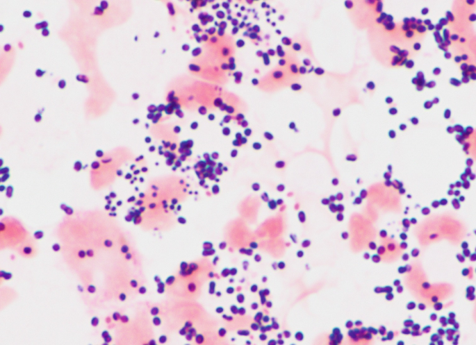
A 9 years old child presented to OPD with complaints of high grade fever, vomiting, one episode of seizure. CSF examination was done and Gram staining of the culture showed the following finding. What is the probable causative agent?
Q14
Flask shaped ulcers in colon are caused by:-
Q15
A neonate was found to have cataract, deafness and cardiac defects. Which group of viruses was the mother infected with?
Q16
Flask-shaped ulcers in the intestine are caused by which of the following?
Q17
A person working in an abattoir presented with malignant pustule on hand. What is the causative agent?
Q18
A 9-year-old child presented to OPD with complaints of high-grade fever, vomiting, and one episode of seizure. CSF examination was done and Gram staining of the culture showed lanceolate-shaped gram-positive diplococci. What is the probable causative agent?
Q19
Rubella is caused by
Q20
A 35 year old man presented with dry cough and rusty colored sputum. He has a history of eating in a Chinese restaurant very often with consumption of crabs. What is the probable causative agent in this condition?